• 文献检索
  • 文档翻译
  • 深度研究
  • 学术资讯
  • Suppr Zotero 插件Zotero 插件
  • 邀请有礼
  • 套餐&价格
  • 历史记录
应用&插件
Suppr Zotero 插件Zotero 插件浏览器插件Mac 客户端Windows 客户端微信小程序
定价
高级版会员购买积分包购买API积分包
服务
文献检索文档翻译深度研究API 文档MCP 服务
关于我们
关于 Suppr公司介绍联系我们用户协议隐私条款
关注我们

Suppr 超能文献

核心技术专利:CN118964589B侵权必究
粤ICP备2023148730 号-1Suppr @ 2026

文献检索

告别复杂PubMed语法,用中文像聊天一样搜索,搜遍4000万医学文献。AI智能推荐,让科研检索更轻松。

立即免费搜索

文件翻译

保留排版,准确专业,支持PDF/Word/PPT等文件格式,支持 12+语言互译。

免费翻译文档

深度研究

AI帮你快速写综述,25分钟生成高质量综述,智能提取关键信息,辅助科研写作。

立即免费体验

非局部颅内腔提取。

Nonlocal intracranial cavity extraction.

作者信息

Manjón José V, Eskildsen Simon F, Coupé Pierrick, Romero José E, Collins D Louis, Robles Montserrat

机构信息

Instituto de Aplicaciones de las Tecnologías de la Información y de las Comunicaciones Avanzadas (ITACA), Universitat Politècnica de València, Camino de Vera s/n, 46022 Valencia, Spain.

Center of Functionally Integrative Neuroscience, Department of Clinical Medicine, Aarhus University, Nørrebrogade 44, 8000 Aarhus, Denmark.

出版信息

Int J Biomed Imaging. 2014;2014:820205. doi: 10.1155/2014/820205. Epub 2014 Sep 28.

DOI:10.1155/2014/820205
PMID:25328511
原文链接:https://pmc.ncbi.nlm.nih.gov/articles/PMC4195262/
Abstract

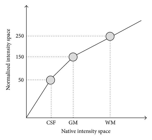
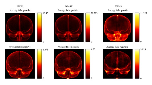
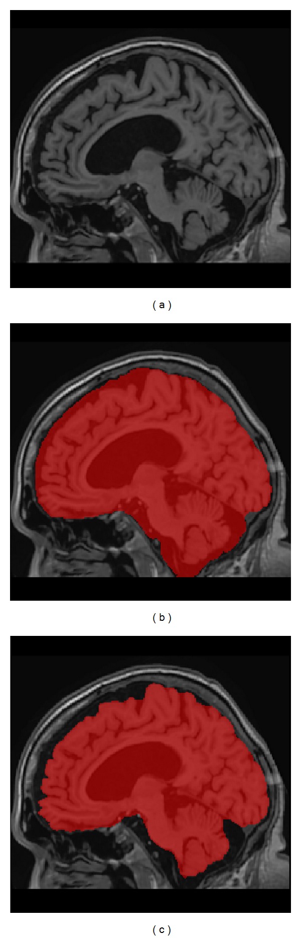
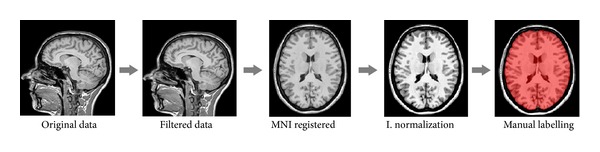
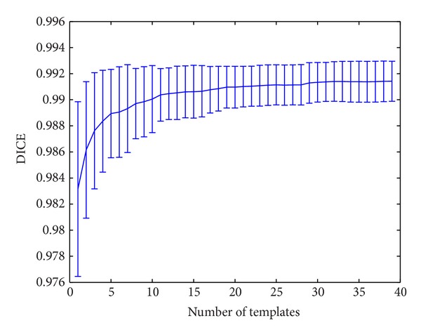
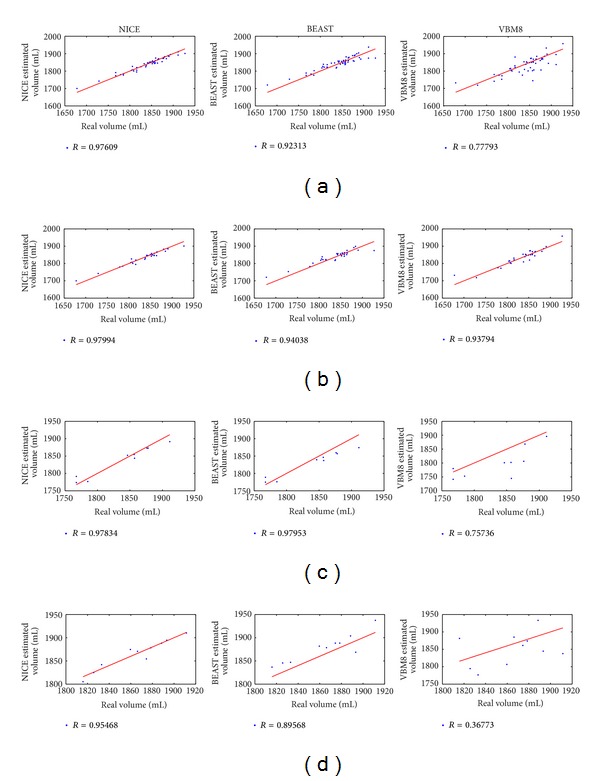
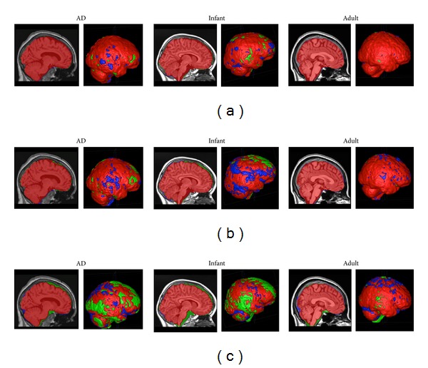
Automatic and accurate methods to estimate normalized regional brain volumes from MRI data are valuable tools which may help to obtain an objective diagnosis and followup of many neurological diseases. To estimate such regional brain volumes, the intracranial cavity volume (ICV) is often used for normalization. However, the high variability of brain shape and size due to normal intersubject variability, normal changes occurring over the lifespan, and abnormal changes due to disease makes the ICV estimation problem challenging. In this paper, we present a new approach to perform ICV extraction based on the use of a library of prelabeled brain images to capture the large variability of brain shapes. To this end, an improved nonlocal label fusion scheme based on BEaST technique is proposed to increase the accuracy of the ICV estimation. The proposed method is compared with recent state-of-the-art methods and the results demonstrate an improved performance both in terms of accuracy and reproducibility while maintaining a reduced computational burden.

摘要

从MRI数据中自动准确估计标准化区域脑容量的方法是有价值的工具,有助于对许多神经系统疾病进行客观诊断和随访。为了估计此类区域脑容量,颅内腔容积(ICV)常被用于标准化。然而,由于个体间正常变异、一生中发生的正常变化以及疾病导致的异常变化,脑形状和大小的高度变异性使得ICV估计问题具有挑战性。在本文中,我们提出了一种新方法,基于使用预标记脑图像库来捕捉脑形状的巨大变异性来进行ICV提取。为此,提出了一种基于BEaST技术的改进非局部标签融合方案,以提高ICV估计的准确性。将所提出的方法与最近的先进方法进行了比较,结果表明,该方法在准确性和可重复性方面均有改进,同时保持了较低的计算负担。

https://cdn.ncbi.nlm.nih.gov/pmc/blobs/4caa/4195262/13c15aab3835/IJBI2014-820205.007.jpg
https://cdn.ncbi.nlm.nih.gov/pmc/blobs/4caa/4195262/cfa043134c6e/IJBI2014-820205.001.jpg
https://cdn.ncbi.nlm.nih.gov/pmc/blobs/4caa/4195262/0c11fe79b010/IJBI2014-820205.002.jpg
https://cdn.ncbi.nlm.nih.gov/pmc/blobs/4caa/4195262/078e9581d784/IJBI2014-820205.003.jpg
https://cdn.ncbi.nlm.nih.gov/pmc/blobs/4caa/4195262/01eefa192d29/IJBI2014-820205.004.jpg
https://cdn.ncbi.nlm.nih.gov/pmc/blobs/4caa/4195262/14ae739a533d/IJBI2014-820205.005.jpg
https://cdn.ncbi.nlm.nih.gov/pmc/blobs/4caa/4195262/990c88bf4e9d/IJBI2014-820205.006.jpg
https://cdn.ncbi.nlm.nih.gov/pmc/blobs/4caa/4195262/13c15aab3835/IJBI2014-820205.007.jpg

相似文献

1
Nonlocal intracranial cavity extraction.非局部颅内腔提取。
Int J Biomed Imaging. 2014;2014:820205. doi: 10.1155/2014/820205. Epub 2014 Sep 28.
2
BEaST: brain extraction based on nonlocal segmentation technique.BEaST:基于非局部分割技术的脑提取。
Neuroimage. 2012 Feb 1;59(3):2362-73. doi: 10.1016/j.neuroimage.2011.09.012. Epub 2011 Sep 16.
3
How Does the Accuracy of Intracranial Volume Measurements Affect Normalized Brain Volumes? Sample Size Estimates Based on 966 Subjects from the HUNT MRI Cohort.颅内体积测量的准确性如何影响标准化脑容量?基于HUNT MRI队列中966名受试者的样本量估计。
AJNR Am J Neuroradiol. 2015 Aug;36(8):1450-6. doi: 10.3174/ajnr.A4299. Epub 2015 Apr 9.
4
The effects of intracranial volume adjustment approaches on multiple regional MRI volumes in healthy aging and Alzheimer's disease.颅内容量调整方法对健康衰老和阿尔茨海默病患者多个脑区 MRI 容量的影响。
Front Aging Neurosci. 2014 Oct 7;6:264. doi: 10.3389/fnagi.2014.00264. eCollection 2014.
5
Intracranial volume estimated with commonly used methods could introduce bias in studies including brain volume measurements.使用常用方法估计的颅内体积可能会在包括脑容量测量的研究中引入偏差。
Neuroimage. 2013 Dec;83:355-60. doi: 10.1016/j.neuroimage.2013.06.068. Epub 2013 Jul 1.
6
A practical guideline for intracranial volume estimation in patients with Alzheimer's disease.阿尔茨海默病患者颅内容量估计的实用指南。
BMC Bioinformatics. 2015;16 Suppl 7(Suppl 7):S8. doi: 10.1186/1471-2105-16-S7-S8. Epub 2015 Apr 23.
7
Validated automatic brain extraction of head CT images.头部CT图像的经验证的自动脑提取
Neuroimage. 2015 Jul 1;114:379-85. doi: 10.1016/j.neuroimage.2015.03.074. Epub 2015 Apr 7.
8
Changes in the intracranial volume from early adulthood to the sixth decade of life: A longitudinal study.从青年到六旬的颅内容积变化:一项纵向研究。
Neuroimage. 2020 Oct 15;220:116842. doi: 10.1016/j.neuroimage.2020.116842. Epub 2020 Apr 24.
9
Estimating Intracranial Volume in Brain Research: An Evaluation of Methods.脑研究中颅内体积的估计:方法评估
Neuroinformatics. 2015 Oct;13(4):427-41. doi: 10.1007/s12021-015-9266-5.
10
Investigating association of brain volumes with intracranial capacity in schizophrenia.探讨精神分裂症脑容量与颅内容量的相关性。
Neuroimage. 2010 Feb 1;49(3):2503-8. doi: 10.1016/j.neuroimage.2009.09.006. Epub 2009 Sep 19.

引用本文的文献

1
Lifespan Tree of Brain Anatomy: Diagnostic Values for Motor and Cognitive Neurodegenerative Diseases.脑解剖学的寿命树:运动和认知神经退行性疾病的诊断价值。
Hum Brain Mapp. 2025 Sep;46(13):e70336. doi: 10.1002/hbm.70336.
2
Intracranial volume segmentation for neurodegenerative populations using multicentre FLAIR MRI.使用多中心液体衰减反转恢复磁共振成像(FLAIR MRI)对神经退行性疾病人群进行颅内体积分割
Neuroimage Rep. 2021 Mar 11;1(1):100006. doi: 10.1016/j.ynirp.2021.100006. eCollection 2021 Mar.
3
MRI Voxel Morphometry Shows Brain Volume Changes in Breast Cancer Survivors: Implications for Treatment.

本文引用的文献

1
Intracranial volume estimated with commonly used methods could introduce bias in studies including brain volume measurements.使用常用方法估计的颅内体积可能会在包括脑容量测量的研究中引入偏差。
Neuroimage. 2013 Dec;83:355-60. doi: 10.1016/j.neuroimage.2013.06.068. Epub 2013 Jul 1.
2
Adjusting for global effects in voxel-based morphometry: gray matter decline in normal aging.基于体素的形态计量学中的全局效应调整:正常衰老中的灰质下降。
Neuroimage. 2012 Apr 2;60(2):1503-16. doi: 10.1016/j.neuroimage.2011.12.086. Epub 2012 Jan 8.
3
BEaST: brain extraction based on nonlocal segmentation technique.
MRI体素形态学显示乳腺癌幸存者的脑容量变化:对治疗的启示。
Pathophysiology. 2025 Mar 12;32(1):11. doi: 10.3390/pathophysiology32010011.
4
Data-driven normative values based on generative manifold learning for quantitative MRI.基于生成流形学习的定量 MRI 数据驱动规范值。
Sci Rep. 2024 Mar 30;14(1):7563. doi: 10.1038/s41598-024-58141-4.
5
Brain structure ages-A new biomarker for multi-disease classification.脑结构老化——多疾病分类的新生物标志物。
Hum Brain Mapp. 2024 Jan;45(1):e26558. doi: 10.1002/hbm.26558.
6
The Cerebellum and Cognitive Function: Anatomical Evidence from a Transdiagnostic Sample.小脑与认知功能:来自跨诊断样本的解剖学证据。
Cerebellum. 2024 Aug;23(4):1399-1410. doi: 10.1007/s12311-023-01645-y. Epub 2023 Dec 27.
7
Deep grading for MRI-based differential diagnosis of Alzheimer's disease and Frontotemporal dementia.基于 MRI 的阿尔茨海默病和额颞叶痴呆的深度分级诊断。
Artif Intell Med. 2023 Oct;144:102636. doi: 10.1016/j.artmed.2023.102636. Epub 2023 Aug 18.
8
Deep Grey Matter Volume is Reduced in Amateur Boxers as Compared to Healthy Age-matched Controls.业余拳击手的大脑深部灰质体积较健康同龄对照组减少。
Clin Neuroradiol. 2023 Jun;33(2):475-482. doi: 10.1007/s00062-022-01233-3. Epub 2022 Dec 16.
9
vol2Brain: A New Online Pipeline for Whole Brain MRI Analysis.vol2Brain:一种用于全脑磁共振成像分析的新型在线流程
Front Neuroinform. 2022 May 24;16:862805. doi: 10.3389/fninf.2022.862805. eCollection 2022.
10
Structural progression of Alzheimer's disease over decades: the MRI staging scheme.阿尔茨海默病数十年的结构进展:MRI分期方案
Brain Commun. 2022 Apr 28;4(3):fcac109. doi: 10.1093/braincomms/fcac109. eCollection 2022.
BEaST:基于非局部分割技术的脑提取。
Neuroimage. 2012 Feb 1;59(3):2362-73. doi: 10.1016/j.neuroimage.2011.09.012. Epub 2011 Sep 16.
4
A comprehensive testing protocol for MRI neuroanatomical segmentation techniques: Evaluation of a novel lateral ventricle segmentation method.用于 MRI 神经解剖分割技术的全面测试方案:新型侧脑室分割方法的评估。
Neuroimage. 2011 Oct 15;58(4):1051-9. doi: 10.1016/j.neuroimage.2011.06.080. Epub 2011 Jul 30.
5
A supervised patch-based approach for human brain labeling.基于监督的斑块方法进行人脑标记。
IEEE Trans Med Imaging. 2011 Oct;30(10):1852-62. doi: 10.1109/TMI.2011.2156806. Epub 2011 May 19.
6
Brain MAPS: an automated, accurate and robust brain extraction technique using a template library.脑图谱:一种基于模板库的自动、准确和稳健的脑提取技术。
Neuroimage. 2011 Apr 1;55(3):1091-108. doi: 10.1016/j.neuroimage.2010.12.067. Epub 2010 Dec 31.
7
Patch-based segmentation using expert priors: application to hippocampus and ventricle segmentation.基于补丁的分割使用专家先验:在海马体和脑室分割中的应用。
Neuroimage. 2011 Jan 15;54(2):940-54. doi: 10.1016/j.neuroimage.2010.09.018. Epub 2010 Sep 17.
8
N4ITK: improved N3 bias correction.N4ITK:改进的 N3 偏置校正。
IEEE Trans Med Imaging. 2010 Jun;29(6):1310-20. doi: 10.1109/TMI.2010.2046908. Epub 2010 Apr 8.
9
A robust method to estimate the intracranial volume across MRI field strengths (1.5T and 3T).一种稳健的方法,用于估计不同 MRI 场强(1.5T 和 3T)下的颅内体积。
Neuroimage. 2010 May 1;50(4):1427-37. doi: 10.1016/j.neuroimage.2010.01.064. Epub 2010 Jan 28.
10
Adaptive non-local means denoising of MR images with spatially varying noise levels.具有空间变化噪声水平的磁共振图像自适应非局部均值去噪
J Magn Reson Imaging. 2010 Jan;31(1):192-203. doi: 10.1002/jmri.22003.