• 文献检索
  • 文档翻译
  • 深度研究
  • 学术资讯
  • Suppr Zotero 插件Zotero 插件
  • 邀请有礼
  • 套餐&价格
  • 历史记录
应用&插件
Suppr Zotero 插件Zotero 插件浏览器插件Mac 客户端Windows 客户端微信小程序
定价
高级版会员购买积分包购买API积分包
服务
文献检索文档翻译深度研究API 文档MCP 服务
关于我们
关于 Suppr公司介绍联系我们用户协议隐私条款
关注我们

Suppr 超能文献

核心技术专利:CN118964589B侵权必究
粤ICP备2023148730 号-1Suppr @ 2026

文献检索

告别复杂PubMed语法,用中文像聊天一样搜索,搜遍4000万医学文献。AI智能推荐,让科研检索更轻松。

立即免费搜索

文件翻译

保留排版,准确专业,支持PDF/Word/PPT等文件格式,支持 12+语言互译。

免费翻译文档

深度研究

AI帮你快速写综述,25分钟生成高质量综述,智能提取关键信息,辅助科研写作。

立即免费体验

一个额外的中间楔骨?

An additional middle cuneiform?

作者信息

Brookes-Fazakerley S D, Jackson G E, Platt S R

机构信息

Department of Trauma & Orthopaedics, Wirral University Teaching Hospital NHS Foundation Trust, Arrowe Park Hospital, Upton, Wirral, Merseyside, UK

Department of Trauma & Orthopaedics, Wirral University Teaching Hospital NHS Foundation Trust, Arrowe Park Hospital, Upton, Wirral, Merseyside, UK.

出版信息

J Surg Case Rep. 2015 Jul 29;2015(7):rjv076. doi: 10.1093/jscr/rjv076.

DOI:10.1093/jscr/rjv076
PMID:26224890
原文链接:https://pmc.ncbi.nlm.nih.gov/articles/PMC4518717/
Abstract

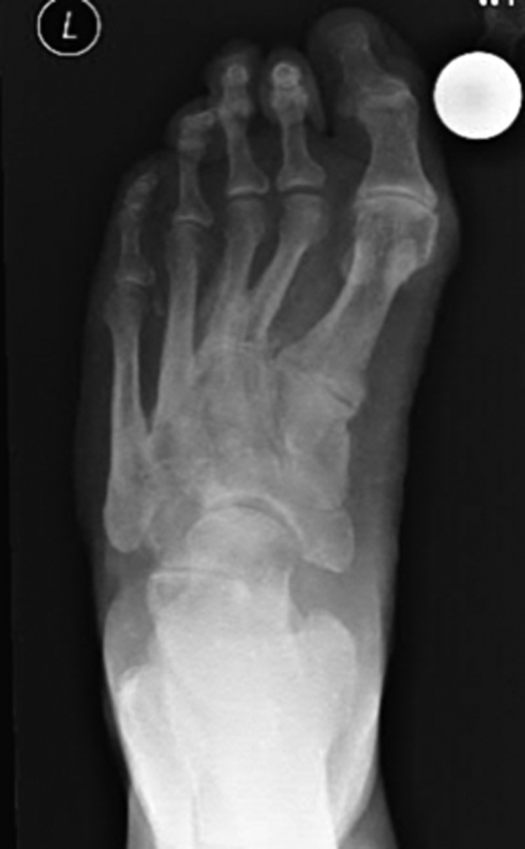
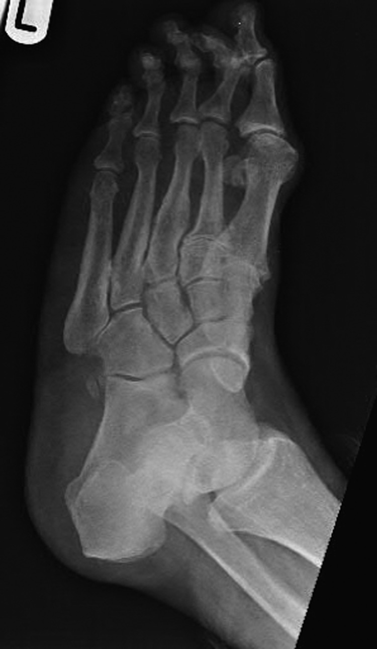
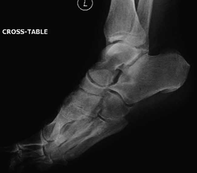
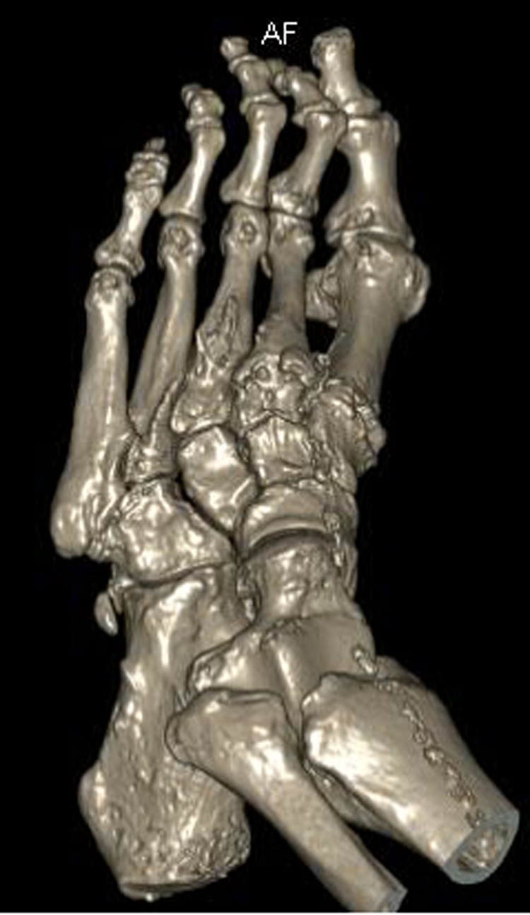
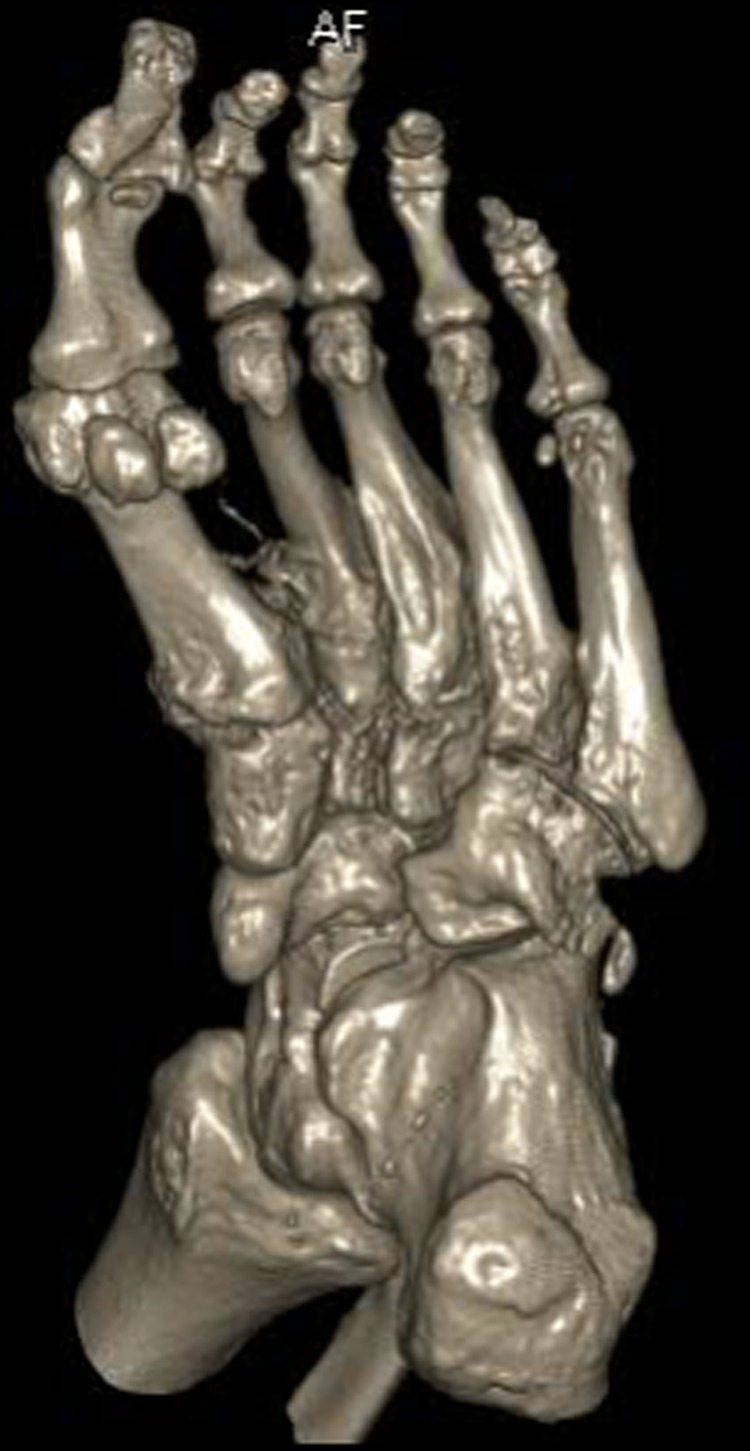
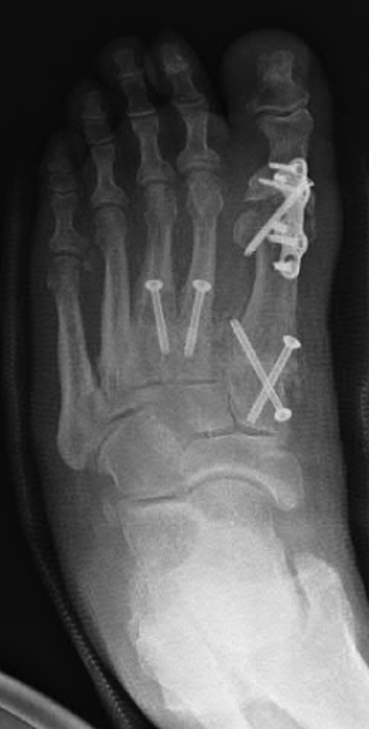
Additional cuneiform bones of the foot have been described in reference to the medial bipartite cuneiform or as small accessory ossicles. An additional middle cuneiform has not been previously documented. We present the case of a patient with an additional ossicle that has the appearance and location of an additional middle cuneiform. Recognizing such an anatomical anomaly is essential for ruling out second metatarsal base or middle cuneiform fractures and for the preoperative planning of arthrodesis or open reduction and internal fixation procedures in this anatomical location.

摘要

足部额外的楔骨已被描述为与内侧二分楔骨相关或作为小的副骨。此前尚未有额外中间楔骨的记录。我们报告一例患者,其有一个外观和位置类似额外中间楔骨的额外小骨。识别这种解剖学异常对于排除第二跖骨基底或中间楔骨骨折以及该解剖部位关节融合术或切开复位内固定手术的术前规划至关重要。

https://cdn.ncbi.nlm.nih.gov/pmc/blobs/6f44/4518717/352af4cdc8c3/rjv07607.jpg
https://cdn.ncbi.nlm.nih.gov/pmc/blobs/6f44/4518717/c101a3a33155/rjv07601.jpg
https://cdn.ncbi.nlm.nih.gov/pmc/blobs/6f44/4518717/55c93fcc1749/rjv07602.jpg
https://cdn.ncbi.nlm.nih.gov/pmc/blobs/6f44/4518717/1a6d020a2008/rjv07603.jpg
https://cdn.ncbi.nlm.nih.gov/pmc/blobs/6f44/4518717/4b81c95ed83e/rjv07604.jpg
https://cdn.ncbi.nlm.nih.gov/pmc/blobs/6f44/4518717/53dec702e303/rjv07605.jpg
https://cdn.ncbi.nlm.nih.gov/pmc/blobs/6f44/4518717/ba8c06b2ddea/rjv07606.jpg
https://cdn.ncbi.nlm.nih.gov/pmc/blobs/6f44/4518717/352af4cdc8c3/rjv07607.jpg

相似文献

1
An additional middle cuneiform?一个额外的中间楔骨?
J Surg Case Rep. 2015 Jul 29;2015(7):rjv076. doi: 10.1093/jscr/rjv076.
2
Bipartite medial cuneiform: magnetic resonance imaging findings and prevalence of this rare anatomical variant.二分体内侧楔骨:这种罕见解剖变异的磁共振成像表现和流行率。
Skeletal Radiol. 2020 May;49(5):691-698. doi: 10.1007/s00256-019-03353-3. Epub 2019 Nov 28.
3
Appropriate Area for Operative Procedures Near Tibialis Anterior Tendon Insertion: A Cadaveric Study.胫骨前肌腱止点附近手术操作的合适区域:一项尸体研究
Foot Ankle Int. 2018 Mar;39(3):349-354. doi: 10.1177/1071100717743964. Epub 2018 Jan 7.
4
[Clinical and radiographic evaluation of open reduction and internal fixation with headless compression screws in treatment of lisfranc joint injuries].无头加压螺钉切开复位内固定治疗Lisfranc关节损伤的临床及影像学评估
Zhongguo Xiu Fu Chong Jian Wai Ke Za Zhi. 2013 Oct;27(10):1196-201.
5
Rate of nonunion after first metatarsal-cuneiform arthrodesis using joint curettage and two crossed compression screw fixation: a systematic review.采用关节刮除术和两枚交叉加压螺钉固定进行第一跖楔关节融合术后的骨不连发生率:一项系统评价
J Foot Ankle Surg. 2011 Nov-Dec;50(6):707-9. doi: 10.1053/j.jfas.2011.08.003. Epub 2011 Sep 10.
6
Magnetic resonance imaging findings in bipartite medial cuneiform - a potential pitfall in diagnosis of midfoot injuries: a case series.二分内侧楔骨的磁共振成像表现——中足损伤诊断中的一个潜在陷阱:病例系列
J Med Case Rep. 2008 Aug 13;2:272. doi: 10.1186/1752-1947-2-272.
7
Observed changes in first metatarsal and medial cuneiform positions after first metatarsophalangeal joint arthrodesis.第一跖趾关节融合术后第一跖骨和内侧楔骨位置的观察性变化。
J Foot Ankle Surg. 2014 Jan-Feb;53(1):32-5. doi: 10.1053/j.jfas.2013.09.011. Epub 2013 Nov 8.
8
Combined medial column primary arthrodesis, middle column open reduction internal fixation, and lateral column pinning for treatment of Lisfranc fracture-dislocation injuries.联合内侧柱一期关节融合术、中柱切开复位内固定术及外侧柱克氏针固定术治疗Lisfranc骨折脱位损伤。
J Foot Ankle Surg. 2014 Sep-Oct;53(5):657-63. doi: 10.1053/j.jfas.2014.04.016. Epub 2014 May 17.
9
In vitro determination of midfoot motion.中足运动的体外测定
Foot Ankle. 1989 Dec;10(3):140-6. doi: 10.1177/107110078901000305.
10
Radiographic Relevance of the Distal Medial Cuneiform Angle in Hallux Valgus Assessment.踇外翻评估中内侧楔骨远端角的影像学相关性
J Foot Ankle Surg. 2016 Jan-Feb;55(1):85-9. doi: 10.1053/j.jfas.2015.06.026. Epub 2015 Sep 7.

引用本文的文献

1
Incidental Detection of Bilateral Tarsal 3rd Middle Phalangeal Bipartition in the Emergency Department: A Case Report.急诊科偶然发现双侧跗骨第三中节指骨二分:一例报告
J Orthop Case Rep. 2024 Dec;14(12):133-136. doi: 10.13107/jocr.2024.v14.i12.5048.
2
Bipartite medial cuneiform: magnetic resonance imaging findings and prevalence of this rare anatomical variant.二分体内侧楔骨:这种罕见解剖变异的磁共振成像表现和流行率。
Skeletal Radiol. 2020 May;49(5):691-698. doi: 10.1007/s00256-019-03353-3. Epub 2019 Nov 28.
3
Anatomical variation in the ankle and foot: from incidental finding to inductor of pathology. Part II: midfooot and forefoot.

本文引用的文献

1
Bipartite medial cuneiform: new frequencies from skeletal collections and a meta-analysis of previous cases.二分内侧楔骨:骨骼样本中的新发生率及既往病例的荟萃分析
Homo. 2011 Apr;62(2):109-25. doi: 10.1016/j.jchb.2011.01.002. Epub 2011 Mar 5.
踝关节和足部的解剖变异:从偶然发现到病理诱导因素。第二部分:中足和前足。
Insights Imaging. 2019 Jul 31;10(1):69. doi: 10.1186/s13244-019-0747-1.