• 文献检索
  • 文档翻译
  • 深度研究
  • 学术资讯
  • Suppr Zotero 插件Zotero 插件
  • 邀请有礼
  • 套餐&价格
  • 历史记录
应用&插件
Suppr Zotero 插件Zotero 插件浏览器插件Mac 客户端Windows 客户端微信小程序
定价
高级版会员购买积分包购买API积分包
服务
文献检索文档翻译深度研究API 文档MCP 服务
关于我们
关于 Suppr公司介绍联系我们用户协议隐私条款
关注我们

Suppr 超能文献

核心技术专利:CN118964589B侵权必究
粤ICP备2023148730 号-1Suppr @ 2026

文献检索

告别复杂PubMed语法,用中文像聊天一样搜索,搜遍4000万医学文献。AI智能推荐,让科研检索更轻松。

立即免费搜索

文件翻译

保留排版,准确专业,支持PDF/Word/PPT等文件格式,支持 12+语言互译。

免费翻译文档

深度研究

AI帮你快速写综述,25分钟生成高质量综述,智能提取关键信息,辅助科研写作。

立即免费体验

踝关节和足部的解剖变异:从偶然发现到病理诱导因素。第二部分:中足和前足。

Anatomical variation in the ankle and foot: from incidental finding to inductor of pathology. Part II: midfooot and forefoot.

作者信息

Aparisi Gómez Maria Pilar, Aparisi Francisco, Bartoloni Alessandra, Ferrando Fons Maria Alejandra, Battista Giuseppe, Guglielmi Giuseppe, Bazzocchi Alberto

机构信息

Department of Radiology, Auckland City Hospital - Auckland District Health Board (ADHB), 2 Park Road, Grafton, Auckland, 1023, New Zealand.

Department of Radiology, Hospital Vithas Nueve de Octubre, Calle Valle de la Ballestera, 59, 46015, Valencia, Spain.

出版信息

Insights Imaging. 2019 Jul 31;10(1):69. doi: 10.1186/s13244-019-0747-1.

DOI:10.1186/s13244-019-0747-1
PMID:31363862
原文链接:https://pmc.ncbi.nlm.nih.gov/articles/PMC6667607/
Abstract

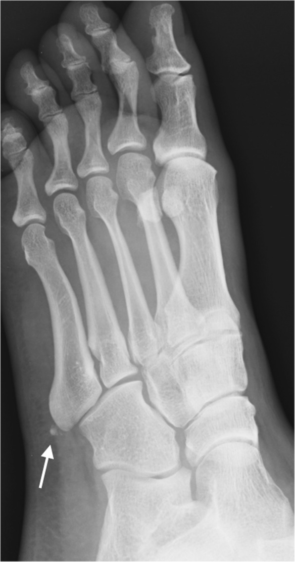
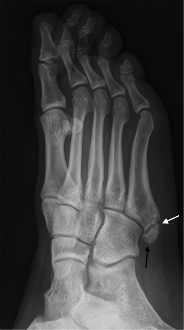
Accessory anatomical structures in the ankle and foot usually represent incidental imaging findings; however, they may also eventually represent a source of pathology, such as painful syndromes, degenerative changes, be the subject of overuse and trauma, or appear as masses and cause compression syndromes or impingement. This review aims to describe and illustrate the imaging findings related to the presence of accessory ossicles and muscles in the midfoot and forefoot through different techniques, with special attention on those variants that associate factors of clinical relevance or that would trigger challenges in the differential diagnosis.

摘要

踝关节和足部的附属解剖结构通常表现为偶然的影像学发现;然而,它们最终也可能成为病理来源,如疼痛综合征、退行性改变、过度使用和创伤的对象,或表现为肿块并导致压迫综合征或撞击。本综述旨在通过不同技术描述和说明与中足和前足附属小骨和肌肉存在相关的影像学发现,特别关注那些与临床相关因素相关或在鉴别诊断中引发挑战的变异。

https://cdn.ncbi.nlm.nih.gov/pmc/blobs/1831/6667607/572e379b3c0c/13244_2019_747_Fig18_HTML.jpg
https://cdn.ncbi.nlm.nih.gov/pmc/blobs/1831/6667607/7378d7479cec/13244_2019_747_Fig1_HTML.jpg
https://cdn.ncbi.nlm.nih.gov/pmc/blobs/1831/6667607/70899080c3d8/13244_2019_747_Fig2_HTML.jpg
https://cdn.ncbi.nlm.nih.gov/pmc/blobs/1831/6667607/1fdecaec4b34/13244_2019_747_Fig3_HTML.jpg
https://cdn.ncbi.nlm.nih.gov/pmc/blobs/1831/6667607/88b3c69517d5/13244_2019_747_Fig4_HTML.jpg
https://cdn.ncbi.nlm.nih.gov/pmc/blobs/1831/6667607/bae7e04781c4/13244_2019_747_Fig5_HTML.jpg
https://cdn.ncbi.nlm.nih.gov/pmc/blobs/1831/6667607/899c0218330c/13244_2019_747_Fig6_HTML.jpg
https://cdn.ncbi.nlm.nih.gov/pmc/blobs/1831/6667607/9fb8954e0394/13244_2019_747_Fig7_HTML.jpg
https://cdn.ncbi.nlm.nih.gov/pmc/blobs/1831/6667607/90f028eebf5b/13244_2019_747_Fig8_HTML.jpg
https://cdn.ncbi.nlm.nih.gov/pmc/blobs/1831/6667607/e6c23d008837/13244_2019_747_Fig9_HTML.jpg
https://cdn.ncbi.nlm.nih.gov/pmc/blobs/1831/6667607/502b25367419/13244_2019_747_Fig10_HTML.jpg
https://cdn.ncbi.nlm.nih.gov/pmc/blobs/1831/6667607/9dd45e1a9818/13244_2019_747_Fig11_HTML.jpg
https://cdn.ncbi.nlm.nih.gov/pmc/blobs/1831/6667607/0b2a161f7a55/13244_2019_747_Fig12_HTML.jpg
https://cdn.ncbi.nlm.nih.gov/pmc/blobs/1831/6667607/4486a31a87ba/13244_2019_747_Fig13_HTML.jpg
https://cdn.ncbi.nlm.nih.gov/pmc/blobs/1831/6667607/a3fa697b7bb6/13244_2019_747_Fig14_HTML.jpg
https://cdn.ncbi.nlm.nih.gov/pmc/blobs/1831/6667607/89ef65016592/13244_2019_747_Fig15_HTML.jpg
https://cdn.ncbi.nlm.nih.gov/pmc/blobs/1831/6667607/18d9c55dd261/13244_2019_747_Fig16_HTML.jpg
https://cdn.ncbi.nlm.nih.gov/pmc/blobs/1831/6667607/1f202b8ed4f5/13244_2019_747_Fig17_HTML.jpg
https://cdn.ncbi.nlm.nih.gov/pmc/blobs/1831/6667607/572e379b3c0c/13244_2019_747_Fig18_HTML.jpg

相似文献

1
Anatomical variation in the ankle and foot: from incidental finding to inductor of pathology. Part II: midfooot and forefoot.踝关节和足部的解剖变异:从偶然发现到病理诱导因素。第二部分:中足和前足。
Insights Imaging. 2019 Jul 31;10(1):69. doi: 10.1186/s13244-019-0747-1.
2
Anatomical variation in the ankle and foot: from incidental finding to inductor of pathology. Part I: ankle and hindfoot.踝关节和足部的解剖变异:从偶然发现到病理诱发因素。第一部分:踝关节和后足。
Insights Imaging. 2019 Jul 31;10(1):74. doi: 10.1186/s13244-019-0746-2.
3
Accessory ossicles of the foot-an imaging conundrum.足部副骨——影像学难题
Emerg Radiol. 2019 Aug;26(4):465-478. doi: 10.1007/s10140-019-01688-x. Epub 2019 Apr 8.
4
Accessory ossicles and sesamoid bones of the ankle and foot: imaging findings, clinical significance and differential diagnosis.踝关节和足部的副骨与籽骨:影像学表现、临床意义及鉴别诊断
Eur Radiol. 2003 Dec;13 Suppl 4:L164-77. doi: 10.1007/s00330-003-2011-8.
5
Accessory ossicles and sesamoid bones of the ankle and foot: imaging findings, clinical significance and differential diagnosis.踝关节和足部的副骨与籽骨:影像学表现、临床意义及鉴别诊断
Eur Radiol. 2003 Dec;13 Suppl 6:L164-77. doi: 10.1007/s00330-003-2011-8. Epub 2003 Aug 6.
6
Sesamoids and accessory ossicles of the foot: anatomical variability and related pathology.足部籽骨和副骨:解剖变异与相关病变。
Insights Imaging. 2013 Oct;4(5):581-93. doi: 10.1007/s13244-013-0277-1. Epub 2013 Sep 5.
7
Symptomatic Accessory Ossicles of the Foot and Ankle.足部和踝关节的有症状副骨。
Curr Probl Diagn Radiol. 2023 Jul-Aug;52(4):300-311. doi: 10.1067/j.cpradiol.2023.03.009. Epub 2023 Mar 23.
8
Imaging of Bone Anatomical Variants Around the Foot and Ankle.足踝周围骨解剖变异的影像学表现
Semin Musculoskelet Radiol. 2023 Apr;27(2):214-220. doi: 10.1055/s-0043-1764224. Epub 2023 Apr 3.
9
Accessory Ossicles of the Foot and Ankle: Disorders and a Review of the Literature.足踝部副骨:疾病及文献综述
Cureus. 2017 Nov 26;9(11):e1881. doi: 10.7759/cureus.1881.
10
Imaging of impingement lesions in the ankle.踝关节撞击性损伤的影像学检查
Top Magn Reson Imaging. 2010 Feb;21(1):15-23. doi: 10.1097/RMR.0b013e31820ef46b.

引用本文的文献

1
Sustainable treatment success of an Os naviculare syndrome using conservative measures, infiltration therapy, and shock waves.采用保守治疗、浸润疗法和冲击波治疗舟状骨综合征取得可持续的治疗成功。
J Surg Case Rep. 2024 Aug 28;2024(8):rjae544. doi: 10.1093/jscr/rjae544. eCollection 2024 Aug.
2
Foot pain in children and adolescents: a problem-based approach in musculoskeletal ultrasonography.儿童和青少年足部疼痛:肌肉骨骼超声检查的基于问题的方法
Ultrasonography. 2024 May;43(3):193-208. doi: 10.14366/usg.24002. Epub 2024 Mar 19.
3
Prevalence and classification of accessory navicular bone: a medical record review.

本文引用的文献

1
Incidence and anatomical variations of accessory navicular bone in patients with foot pain: A retrospective radiographic analysis.足痛患者副舟骨的发生率及解剖变异:一项回顾性影像学分析。
Clin Anat. 2017 May;30(4):436-444. doi: 10.1002/ca.22876. Epub 2017 Apr 3.
2
Os peroneum imaging: normal appearance and pathological findings.腓籽骨成像:正常表现及病理结果
Insights Imaging. 2017 Feb;8(1):59-68. doi: 10.1007/s13244-016-0540-3. Epub 2017 Jan 5.
3
Peroneal Tendon Disorders.腓骨肌腱疾病
副舟骨的患病率和分类:病历回顾。
Ann Saudi Med. 2022 Sep-Oct;42(5):327-333. doi: 10.5144/0256-4947.2022.327. Epub 2022 Oct 6.
Clin Sports Med. 2015 Oct;34(4):625-41. doi: 10.1016/j.csm.2015.06.003. Epub 2015 Jul 31.
4
An additional middle cuneiform?一个额外的中间楔骨?
J Surg Case Rep. 2015 Jul 29;2015(7):rjv076. doi: 10.1093/jscr/rjv076.
5
Os intermetatarseum - A case report.跖间骨——一例报告。
J Clin Orthop Trauma. 2012 Jun;3(1):54-7. doi: 10.1016/j.jcot.2011.11.002. Epub 2012 Jun 16.
6
Accessory ossicles and sesamoid bones: recognition and treatment.副骨与籽骨:识别与治疗
Emerg Nurse. 2015 Mar;22(10):27-32. doi: 10.7748/en.22.10.27.e1404.
7
The os vesalianum: an unusual cause of lateral foot pain a case report and review of the literature.维萨里骨:足外侧疼痛的罕见病因——病例报告及文献综述
Orthopedics. 1984 Jan 1;7(1):86-9. doi: 10.3928/0147-7447-19840101-12.
8
Magnetic resonance imaging in adolescent symptomatic navicular tuberosity.青少年有症状的舟骨粗隆的磁共振成像
J Med Invest. 2014;61(1-2):22-7. doi: 10.2152/jmi.61.22.
9
Imaging of tarsal navicular disorders: a pictorial review.跗舟骨疾病的影像学:图文综述
Foot Ankle Spec. 2014 Jun;7(3):211-25. doi: 10.1177/1938640014528042. Epub 2014 Mar 30.
10
Sesamoids and accessory ossicles of the foot: anatomical variability and related pathology.足部籽骨和副骨:解剖变异与相关病变。
Insights Imaging. 2013 Oct;4(5):581-93. doi: 10.1007/s13244-013-0277-1. Epub 2013 Sep 5.