Sousa Sofia, Brion Régis, Lintunen Minnamaija, Kronqvist Pauliina, Sandholm Jouko, Mönkkönen Jukka, Kellokumpu-Lehtinen Pirkko-Liisa, Lauttia Susanna, Tynninen Olli, Joensuu Heikki, Heymann Dominique, Määttä Jorma A
School of Pharmacy, Faculty of Health Sciences, University of Eastern Finland, Yliopistonranta 1C, P.O. Box 1627, FI-70211, Kuopio, Finland.
INSERM, UMR957, Equipe LIGUE 2012, Nantes, F-44035, France.
Breast Cancer Res. 2015 Aug 5;17(1):101. doi: 10.1186/s13058-015-0621-0.
The immune system plays a major role in cancer progression. In solid tumors, 5-40 % of the tumor mass consists of tumor-associated macrophages (TAMs) and there is usually a correlation between the number of TAMs and poor prognosis, depending on the tumor type. TAMs usually resemble M2 macrophages. Unlike M1-macrophages which have pro-inflammatory and anti-cancer functions, M2-macrophages are immunosuppressive, contribute to the matrix-remodeling, and hence favor tumor growth. The role of TAMs is not fully understood in breast cancer progression.
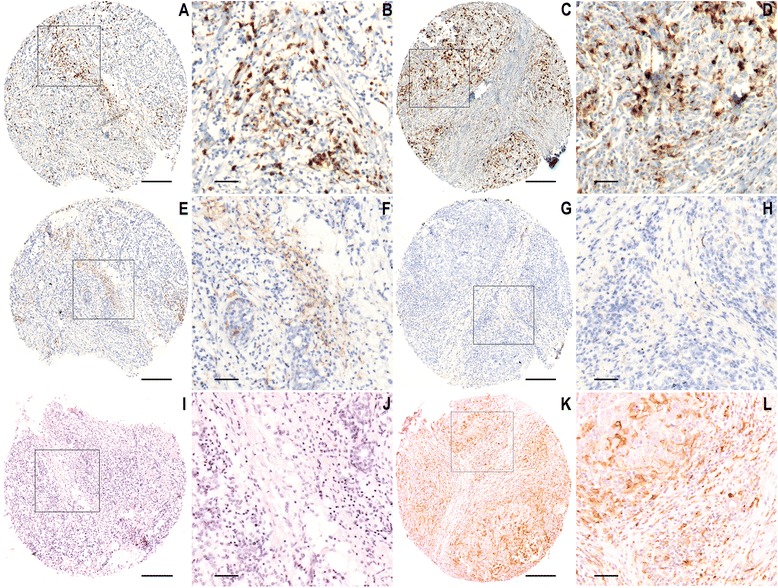
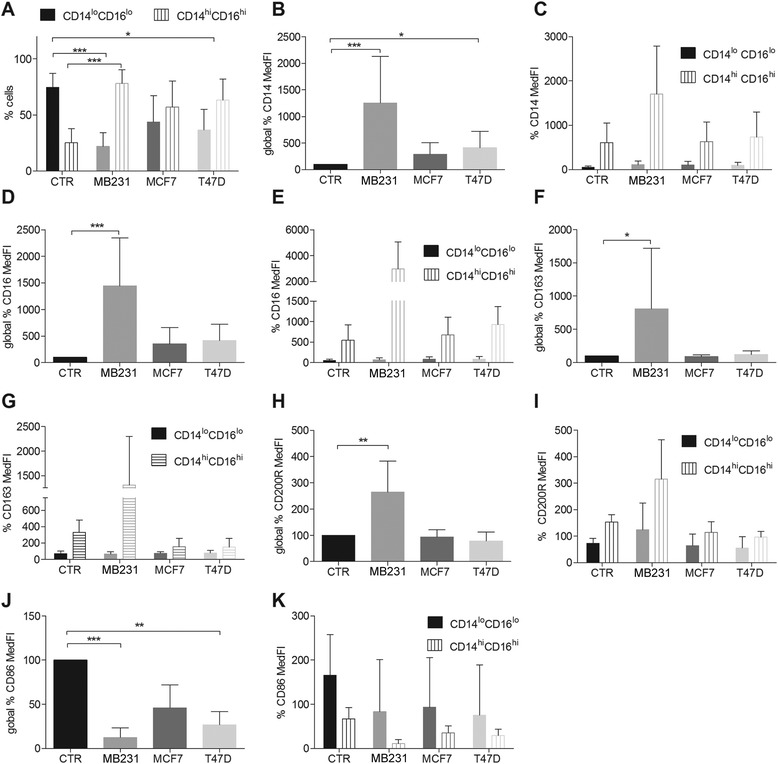
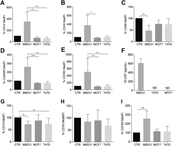
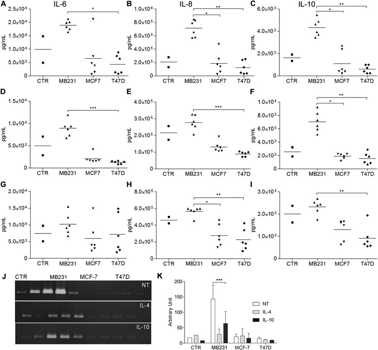
Macrophage infiltration (CD68) and activation status (HLA-DRIIα, CD163) were evaluated in a large cohort of human primary breast tumors (562 tissue microarray samples), by immunohistochemistry and scored by automated image analysis algorithms. Survival between groups was compared using the Kaplan-Meier life-table method and a Cox multivariate proportional hazards model. Macrophage education by breast cancer cells was assessed by ex vivo differentiation of peripheral blood mononuclear cells (PBMCs) in the presence or absence of breast cancer cell conditioned media (MDA-MB231, MCF-7 or T47D cell lines) and M1 or M2 inducing cytokines (respectively IFN-γ, IL-4 and IL-10). Obtained macrophages were analyzed by flow cytometry (CD14, CD16, CD64, CD86, CD200R and CD163), ELISA (IL-6, IL-8, IL-10, monocyte colony stimulating factor M-CSF) and zymography (matrix metalloproteinase 9, MMP-9).
Clinically, we found that high numbers of CD163(+) M2-macrophages were strongly associated with fast proliferation, poor differentiation, estrogen receptor negativity and histological ductal type (p<0.001) in the studied cohort of human primary breast tumors. We demonstrated ex vivo that breast cancer cell-secreted factors modulate macrophage differentiation toward the M2 phenotype. Furthermore, the more aggressive mesenchymal-like cell line MDA-MB231, which secretes high levels of M-CSF, skews macrophages toward the more immunosuppressive M2c subtype.
This study demonstrates that human breast cancer cells influence macrophage differentiation and that TAM differentiation status correlates with recurrence free survival, thus further emphasizing that TAMs can similarly affect therapy efficacy and patient outcome.
免疫系统在癌症进展中起主要作用。在实体瘤中,5% - 40%的肿瘤组织由肿瘤相关巨噬细胞(TAM)组成,并且根据肿瘤类型,TAM的数量与预后不良通常存在相关性。TAM通常类似于M2巨噬细胞。与具有促炎和抗癌功能的M1巨噬细胞不同,M2巨噬细胞具有免疫抑制作用,有助于基质重塑,因此有利于肿瘤生长。TAM在乳腺癌进展中的作用尚未完全明确。
通过免疫组织化学对一大群人原发性乳腺肿瘤(562个组织微阵列样本)中的巨噬细胞浸润(CD68)和活化状态(HLA - DRIIα、CD163)进行评估,并通过自动图像分析算法进行评分。使用Kaplan - Meier生命表法和Cox多变量比例风险模型比较组间生存率。通过在存在或不存在乳腺癌细胞条件培养基(MDA - MB231、MCF - 7或T47D细胞系)以及M1或M2诱导细胞因子(分别为IFN - γ、IL - 4和IL - 10)的情况下对外周血单核细胞(PBMC)进行体外分化,评估乳腺癌细胞对巨噬细胞的诱导作用。通过流式细胞术(CD14、CD16、CD64、CD86、CD200R和CD163)、ELISA(IL - 6、IL - 8、IL - 10、单核细胞集落刺激因子M - CSF)和酶谱分析(基质金属蛋白酶-9,MMP - 9)对获得的巨噬细胞进行分析。
临床上,我们发现在所研究的人原发性乳腺肿瘤队列中,大量CD163(+) M2巨噬细胞与快速增殖、低分化、雌激素受体阴性和组织学导管类型密切相关(p<0.001)。我们在体外证明,乳腺癌细胞分泌的因子可调节巨噬细胞向M2表型分化。此外,分泌高水平M - CSF的更具侵袭性的间充质样细胞系MDA - MB231使巨噬细胞向更具免疫抑制性的M2c亚型倾斜。
本研究表明人乳腺癌细胞会影响巨噬细胞分化,且TAM分化状态与无复发生存率相关,从而进一步强调TAM同样会影响治疗效果和患者预后。