• 文献检索
  • 文档翻译
  • 深度研究
  • 学术资讯
  • Suppr Zotero 插件Zotero 插件
  • 邀请有礼
  • 套餐&价格
  • 历史记录
应用&插件
Suppr Zotero 插件Zotero 插件浏览器插件Mac 客户端Windows 客户端微信小程序
定价
高级版会员购买积分包购买API积分包
服务
文献检索文档翻译深度研究API 文档MCP 服务
关于我们
关于 Suppr公司介绍联系我们用户协议隐私条款
关注我们

Suppr 超能文献

核心技术专利:CN118964589B侵权必究
粤ICP备2023148730 号-1Suppr @ 2026

文献检索

告别复杂PubMed语法,用中文像聊天一样搜索,搜遍4000万医学文献。AI智能推荐,让科研检索更轻松。

立即免费搜索

文件翻译

保留排版,准确专业,支持PDF/Word/PPT等文件格式,支持 12+语言互译。

免费翻译文档

深度研究

AI帮你快速写综述,25分钟生成高质量综述,智能提取关键信息,辅助科研写作。

立即免费体验

使用屏气 BOLD fMRI 测量脑胶质瘤患者的血管反应性图

Vascular Reactivity Maps in Patients with Gliomas Using Breath-Holding BOLD fMRI.

出版信息

J Neuroimaging. 2016 Mar-Apr;26(2):232-9. doi: 10.1111/jon.12278.

DOI:10.1111/jon.12278
PMID:26250554
原文链接:https://pmc.ncbi.nlm.nih.gov/articles/PMC5087143/
Abstract

BACKGROUND AND PURPOSE

To evaluate whether breath-holding (BH) blood oxygenation level-dependent (BOLD) fMRI can quantify differences in vascular reactivity (VR), as there is a need for improved contrast mechanisms in gliomas.

METHODS

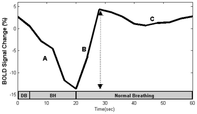
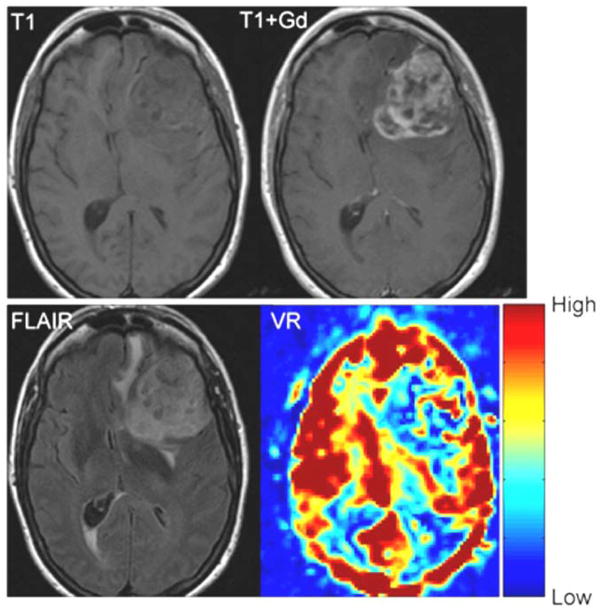
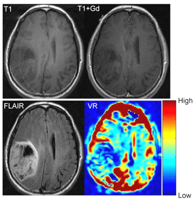
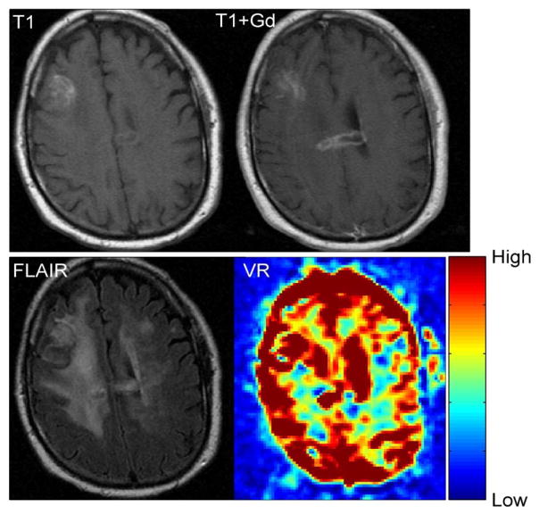
16 patients (gliomas, grade II = 5, III = 2, IV = 9) were evaluated using the BH paradigm: 4-second single deep breath followed by 16 seconds of BH and 40 seconds of regular breathing for five cycles. VR was defined as the difference in BOLD signal between the minimal signal seen at the end of the deep breath and maximal signal seen at the end of BH (peak-to-trough). VR was measured for every voxel and compared for gray versus white matter and tumor versus normal contralateral brain. VR maps were compared to the areas of enhancement and FLAIR/T2 abnormality.

RESULTS

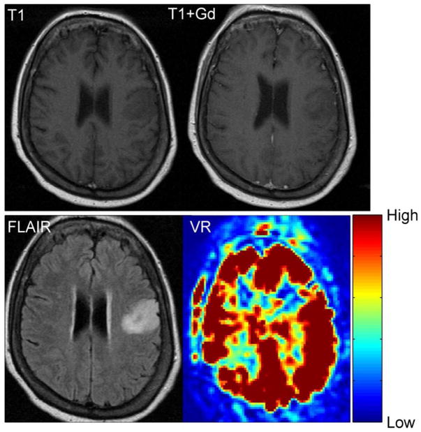
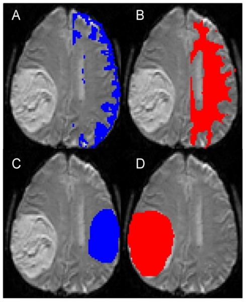
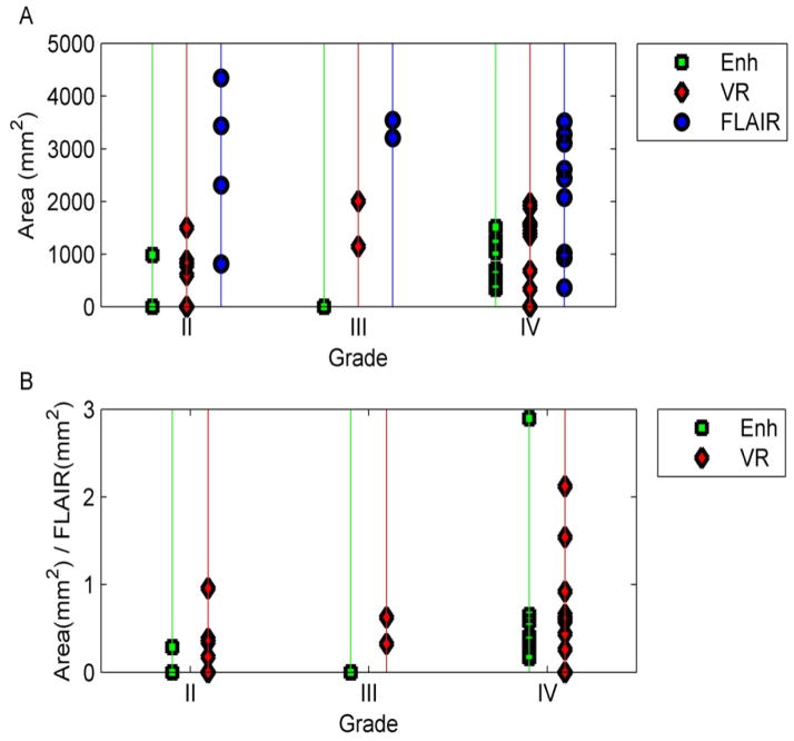
VR was significantly lower in normal white matter than gray matter (P < .05) and in tumors compared to the normal, contralateral brain (P < 0.002). The area of abnormal VR (1103 ± 659 mm²) was significantly greater (P = .019) than the enhancement (543 ± 530 mm²), but significantly smaller (P = .0011) than the FLAIR abnormality (2363 ± 1232 mm²). However, the variability in the areas of gadolinium contrast enhancement versus VR abnormality indicates that the contrast mechanism elicited by BH (caused by abnormal arteriolar smooth muscles) appears to be fundamentally different from the contrast mechanism of gadolinium enhancement (caused by the presence of "leaky" gap junctions).

CONCLUSIONS

BH maps based on peak-to-trough can be used to characterize VR in brain tumors. VR maps in brain tumor patients appear to be caused by a different mechanism than gadolinium enhancement.

摘要

背景与目的

评估屏气(BH)血氧水平依赖(BOLD) fMRI 是否可定量测量血管反应性(VR)的差异,因为需要改善脑肿瘤的对比机制。

方法

16 例患者(脑胶质瘤,2 级=5 例,3 级=2 例,4 级=9 例)接受 BH 范式评估:4 秒深吸气后进行 16 秒 BH 和 40 秒常规呼吸,共 5 个周期。VR 定义为在深呼气结束时看到的最小信号与 BH 结束时看到的最大信号(峰-谷)之间的 BOLD 信号差异。为每个体素测量 VR,并比较灰白质和肿瘤与对侧正常脑之间的差异。VR 图与增强和 FLAIR/T2 异常区域进行比较。

结果

正常白质的 VR 明显低于灰质(P<0.05),肿瘤的 VR 明显低于对侧正常脑(P<0.002)。异常 VR 区域(1103±659 mm²)明显大于增强区域(543±530 mm²)(P=0.019),但明显小于 FLAIR 异常区域(2363±1232 mm²)(P=0.0011)。然而,钆对比增强与 VR 异常区域的面积变异性表明,BH 引起的对比机制(由异常的小动脉平滑肌引起)似乎与钆增强的对比机制(由“渗漏”的缝隙连接的存在引起)在根本上不同。

结论

基于峰-谷的 BH 图可用于表征脑肿瘤的 VR。脑肿瘤患者的 VR 图似乎是由不同于钆增强的机制引起的。

https://cdn.ncbi.nlm.nih.gov/pmc/blobs/2448/5087143/d80293c6798c/nihms822308f8.jpg
https://cdn.ncbi.nlm.nih.gov/pmc/blobs/2448/5087143/64c645c8896b/nihms822308f1.jpg
https://cdn.ncbi.nlm.nih.gov/pmc/blobs/2448/5087143/8be5f15aea5a/nihms822308f2.jpg
https://cdn.ncbi.nlm.nih.gov/pmc/blobs/2448/5087143/29cc0bdbf3d6/nihms822308f3.jpg
https://cdn.ncbi.nlm.nih.gov/pmc/blobs/2448/5087143/f061c65547e0/nihms822308f4.jpg
https://cdn.ncbi.nlm.nih.gov/pmc/blobs/2448/5087143/c5e9fddd1aea/nihms822308f5.jpg
https://cdn.ncbi.nlm.nih.gov/pmc/blobs/2448/5087143/f24cd0d4487b/nihms822308f6.jpg
https://cdn.ncbi.nlm.nih.gov/pmc/blobs/2448/5087143/3b2192028f68/nihms822308f7.jpg
https://cdn.ncbi.nlm.nih.gov/pmc/blobs/2448/5087143/d80293c6798c/nihms822308f8.jpg

相似文献

1
Vascular Reactivity Maps in Patients with Gliomas Using Breath-Holding BOLD fMRI.使用屏气 BOLD fMRI 测量脑胶质瘤患者的血管反应性图
J Neuroimaging. 2016 Mar-Apr;26(2):232-9. doi: 10.1111/jon.12278.
2
Comparison of BOLD cerebrovascular reactivity mapping and DSC MR perfusion imaging for prediction of neurovascular uncoupling potential in brain tumors.BOLD 脑血管反应性 mapping 与 DSC MR 灌注成像在脑肿瘤中预测神经血管脱耦联潜能的比较。
Technol Cancer Res Treat. 2012 Aug;11(4):361-74. doi: 10.7785/tcrt.2012.500284. Epub 2012 Mar 1.
3
Cerebrovascular reactivity mapping in patients with low grade gliomas undergoing presurgical sensorimotor mapping with BOLD fMRI.采用血氧水平依赖性功能磁共振成像(BOLD fMRI)对低级别胶质瘤患者进行术前感觉运动功能区映射时的脑血管反应性图谱分析。
J Magn Reson Imaging. 2014 Aug;40(2):383-90. doi: 10.1002/jmri.24406. Epub 2013 Nov 4.
4
Diffuse gliomas exhibit whole brain impaired cerebrovascular reactivity.弥漫性胶质瘤表现出全脑脑血管反应性受损。
Magn Reson Imaging. 2018 Jan;45:78-83. doi: 10.1016/j.mri.2017.09.017. Epub 2017 Oct 3.
5
Re-examine tumor-induced alterations in hemodynamic responses of BOLD fMRI: implications in presurgical brain mapping.重新审视肿瘤引起的血氧水平依赖性功能磁共振成像血流动力学反应变化:对术前脑图谱绘制的意义。
Acta Radiol. 2012 Sep 1;53(7):802-11. doi: 10.1258/ar.2012.120118. Epub 2012 Jul 31.
6
Using relative cerebral blood flow and volume to evaluate the histopathologic grade of cerebral gliomas: preliminary results.利用相对脑血流量和体积评估脑胶质瘤的组织病理学分级:初步结果
AJR Am J Roentgenol. 2002 Sep;179(3):783-9. doi: 10.2214/ajr.179.3.1790783.
7
Neurovascular uncoupling in resting state fMRI demonstrated in patients with primary brain gliomas.原发性脑胶质瘤患者静息态功能磁共振成像中神经血管解耦的表现
J Magn Reson Imaging. 2016 Mar;43(3):620-6. doi: 10.1002/jmri.25012. Epub 2015 Jul 22.
8
ICA-based denoising strategies in breath-hold induced cerebrovascular reactivity mapping with multi echo BOLD fMRI.基于 ICA 的降噪策略在多回波 BOLD fMRI 屏气诱导脑血管反应性映射中的应用。
Neuroimage. 2021 Jun;233:117914. doi: 10.1016/j.neuroimage.2021.117914. Epub 2021 Mar 6.
9
Blood oxygenation level-dependent MRI of cerebral gliomas during breath holding.屏气期间脑胶质瘤的血氧水平依赖性功能磁共振成像
J Magn Reson Imaging. 2004 Feb;19(2):160-7. doi: 10.1002/jmri.10447.
10
Fourier modeling of the BOLD response to a breath-hold task: Optimization and reproducibility.对屏气任务的血氧水平依赖(BOLD)反应的傅里叶建模:优化与可重复性。
Neuroimage. 2016 Jul 15;135:223-31. doi: 10.1016/j.neuroimage.2016.02.037. Epub 2016 Feb 22.

引用本文的文献

1
Research Progress on Glioma Microenvironment and Invasiveness Utilizing Advanced Multi-Parametric Quantitative MRI.利用先进的多参数定量磁共振成像对胶质瘤微环境与侵袭性的研究进展
Cancers (Basel). 2024 Dec 29;17(1):74. doi: 10.3390/cancers17010074.
2
Estimation of cerebrovascular reactivity amplitude and lag using breath-holding fMRI and the global BOLD signal: Application in diabetes and hypertension.利用屏气功能磁共振成像和全脑血氧水平依赖信号估计脑血管反应性幅度和延迟:在糖尿病和高血压中的应用
J Cereb Blood Flow Metab. 2025 Mar;45(3):459-475. doi: 10.1177/0271678X241270420. Epub 2024 Sep 3.
3
Recommendations for quantitative cerebral perfusion MRI using multi-timepoint arterial spin labeling: Acquisition, quantification, and clinical applications.

本文引用的文献

1
Diffusion and perfusion MRI to differentiate treatment-related changes including pseudoprogression from recurrent tumors in high-grade gliomas with histopathologic evidence.采用扩散加权磁共振成像(Diffusion MRI)和灌注加权磁共振成像(perfusion MRI)鉴别高级别胶质瘤治疗相关改变,包括与肿瘤复发相鉴别的假性进展,并提供组织病理学证据。
AJNR Am J Neuroradiol. 2015 May;36(5):877-85. doi: 10.3174/ajnr.A4218. Epub 2015 Jan 15.
2
Which combination of MR imaging modalities is best for predicting recurrent glioblastoma? Study of diagnostic accuracy and reproducibility.哪种磁共振成像方式组合最适合预测复发性脑胶质瘤?诊断准确性和可重复性研究。
Radiology. 2014 Dec;273(3):831-43. doi: 10.1148/radiol.14132868. Epub 2014 May 30.
3
多时间点动脉自旋标记定量脑灌注 MRI 推荐:采集、定量和临床应用。
Magn Reson Med. 2024 Aug;92(2):469-495. doi: 10.1002/mrm.30091. Epub 2024 Apr 9.
4
Detection and Mitigation of Neurovascular Uncoupling in Brain Gliomas.脑胶质瘤中神经血管解偶联的检测与缓解
Cancers (Basel). 2023 Sep 8;15(18):4473. doi: 10.3390/cancers15184473.
5
fMRI studies evaluating central respiratory control in humans.评估人类中枢呼吸控制的 fMRI 研究。
Front Neural Circuits. 2022 Sep 23;16:982963. doi: 10.3389/fncir.2022.982963. eCollection 2022.
6
Hemodynamic Imaging in Cerebral Diffuse Glioma-Part B: Molecular Correlates, Treatment Effect Monitoring, Prognosis, and Future Directions.脑弥漫性胶质瘤的血流动力学成像 - 第二部分:分子关联、治疗效果监测、预后及未来方向
Cancers (Basel). 2022 Mar 5;14(5):1342. doi: 10.3390/cancers14051342.
7
Feasibility of glioblastoma tissue response mapping with physiologic BOLD imaging using precise oxygen and carbon dioxide challenge.利用精确的氧和二氧化碳挑战进行生理 BOLD 成像对脑胶质母细胞瘤组织反应进行测绘的可行性
MAGMA. 2022 Feb;35(1):29-44. doi: 10.1007/s10334-021-00980-7. Epub 2021 Dec 7.
8
Effects of Thresholding on Voxel-Wise Correspondence of Breath-Hold and Resting-State Maps of Cerebrovascular Reactivity.阈值处理对脑血管反应性屏气和静息态图谱体素对应关系的影响。
Front Neurosci. 2021 Aug 24;15:654957. doi: 10.3389/fnins.2021.654957. eCollection 2021.
9
BOLD asynchrony elucidates tumor burden in IDH-mutated gliomas.BOLD 异步凸显 IDH 突变型脑胶质瘤的肿瘤负荷。
Neuro Oncol. 2022 Jan 5;24(1):78-87. doi: 10.1093/neuonc/noab154.
10
A practical modification to a resting state fMRI protocol for improved characterization of cerebrovascular function.一种改进静息态 fMRI 协议以改善脑血管功能表征的实用方法。
Neuroimage. 2021 Oct 1;239:118306. doi: 10.1016/j.neuroimage.2021.118306. Epub 2021 Jun 24.
Cerebrovascular reactivity mapping in patients with low grade gliomas undergoing presurgical sensorimotor mapping with BOLD fMRI.
采用血氧水平依赖性功能磁共振成像(BOLD fMRI)对低级别胶质瘤患者进行术前感觉运动功能区映射时的脑血管反应性图谱分析。
J Magn Reson Imaging. 2014 Aug;40(2):383-90. doi: 10.1002/jmri.24406. Epub 2013 Nov 4.
4
Three-dimensional amide proton transfer MR imaging of gliomas: Initial experience and comparison with gadolinium enhancement.脑胶质瘤三维酰胺质子转移磁共振成像:初步经验及与钆增强对比
J Magn Reson Imaging. 2013 Nov;38(5):1119-28. doi: 10.1002/jmri.24067. Epub 2013 Feb 25.
5
MRI perfusion in determining pseudoprogression in patients with glioblastoma.磁共振灌注成像在胶质母细胞瘤患者假性进展中的应用。
Clin Imaging. 2013 Jan-Feb;37(1):41-9. doi: 10.1016/j.clinimag.2012.02.016. Epub 2012 Jun 8.
6
Comparison of BOLD cerebrovascular reactivity mapping and DSC MR perfusion imaging for prediction of neurovascular uncoupling potential in brain tumors.BOLD 脑血管反应性 mapping 与 DSC MR 灌注成像在脑肿瘤中预测神经血管脱耦联潜能的比较。
Technol Cancer Res Treat. 2012 Aug;11(4):361-74. doi: 10.7785/tcrt.2012.500284. Epub 2012 Mar 1.
7
Clinical utility of cerebrovascular reactivity mapping in patients with low grade gliomas.低级别胶质瘤患者脑血管反应性图谱的临床应用价值
World J Clin Oncol. 2011 Dec 10;2(12):397-403. doi: 10.5306/wjco.v2.i12.397.
8
Potential utility of conventional MRI signs in diagnosing pseudoprogression in glioblastoma.常规 MRI 征象在诊断胶质母细胞瘤假性进展中的潜在作用。
Neurology. 2011 May 31;76(22):1918-24. doi: 10.1212/WNL.0b013e31821d74e7.
9
Annual report to the nation on the status of cancer, 1975-2007, featuring tumors of the brain and other nervous system.国家癌症报告:1975-2007 年癌症发病与死亡统计,重点介绍脑和神经系统肿瘤。
J Natl Cancer Inst. 2011 May 4;103(9):714-36. doi: 10.1093/jnci/djr077. Epub 2011 Mar 31.
10
The paradoxical effect of bevacizumab in the therapy of malignant gliomas.贝伐珠单抗治疗恶性脑胶质瘤的矛盾效应。
Neurology. 2011 Jan 4;76(1):87-93. doi: 10.1212/WNL.0b013e318204a3af.