Batson Melissa A, Petridou Natalia, Klomp Dennis W J, Frens Maarten A, Neggers Sebastiaan F W
Brain Center Rudolf Magnus, Department of Psychiatry, University Medical Center Utrecht, Utrecht, The Netherlands; Department of Neuroscience, Erasmus MC, Rotterdam, The Netherlands.
Radiology Department, Imaging Division, University Medical Center Utrecht, Utrecht, The Netherlands.
PLoS One. 2015 Aug 10;10(8):e0134933. doi: 10.1371/journal.pone.0134933. eCollection 2015.
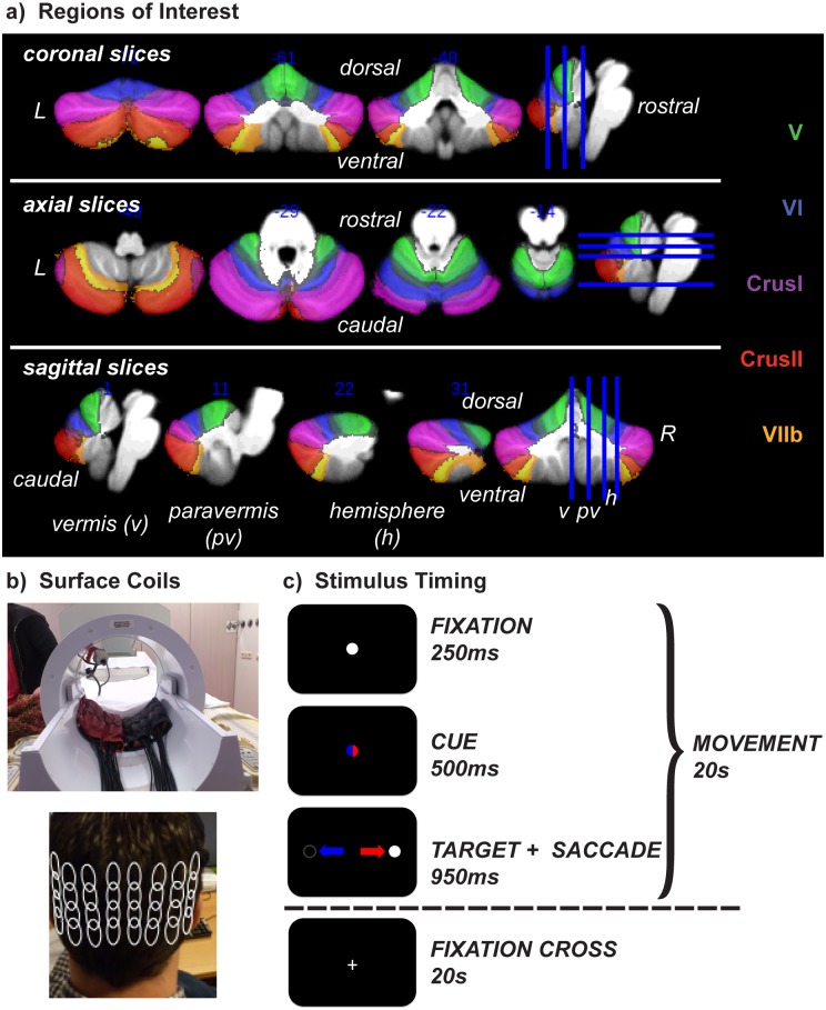
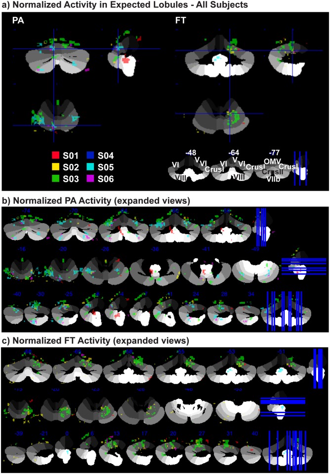
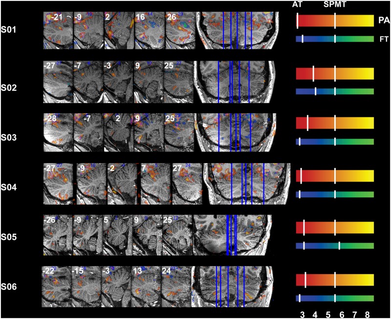
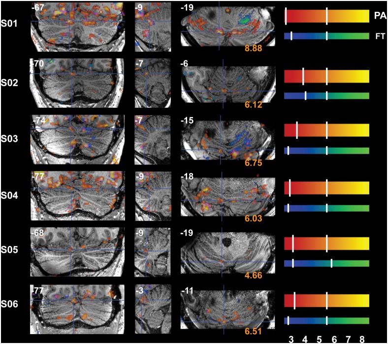
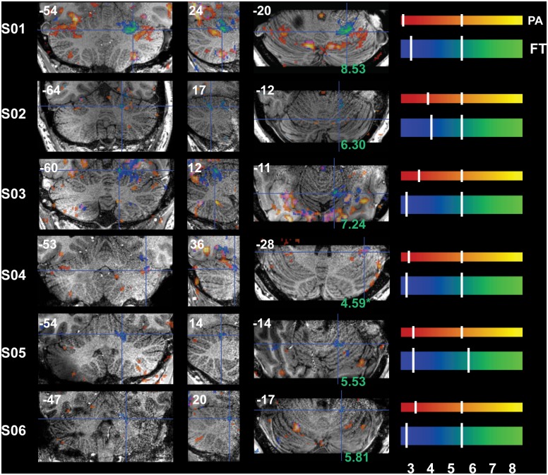
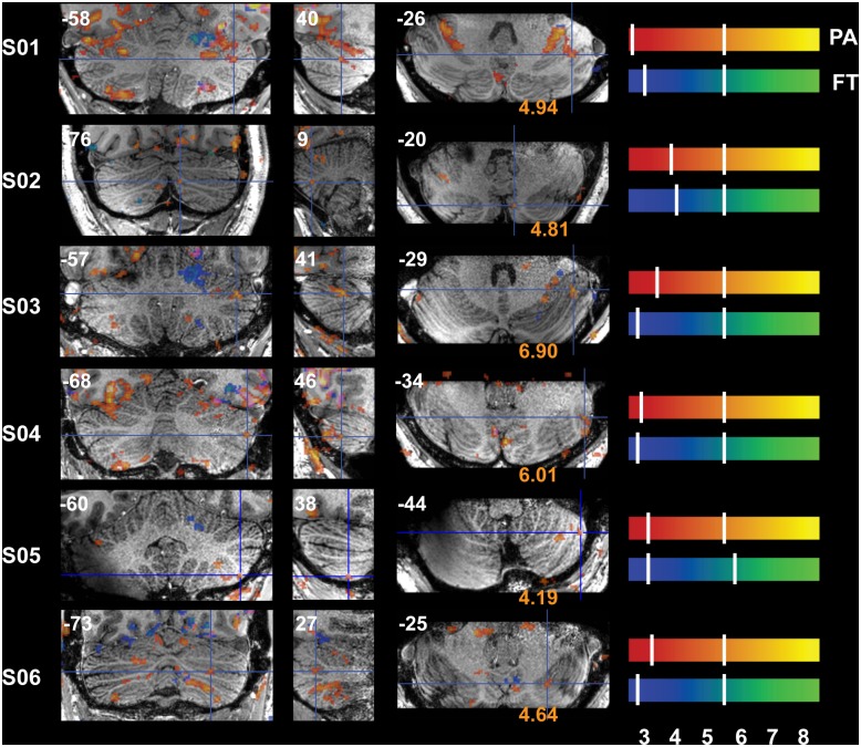
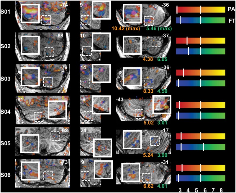
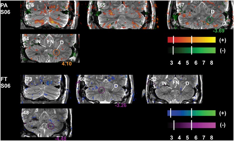
The recent increase in the use of high field MR systems is accompanied by a demand for acquisition techniques and coil systems that can take advantage of increased power and accuracy without being susceptible to increased noise. Physical location and anatomical complexity of targeted regions must be considered when attempting to image deeper structures with small nuclei and/or complex cytoarchitechtonics (i.e. small microvasculature and deep nuclei), such as the brainstem and the cerebellum (Cb). Once these obstacles are overcome, the concomitant increase in signal strength at higher field strength should allow for faster acquisition of MR images. Here we show that it is technically feasible to quickly and accurately detect blood oxygen level dependent (BOLD) signal changes and obtain anatomical images of Cb at high spatial resolutions in individual subjects at 7 Tesla in a single one-hour session. Images were obtained using two high-density multi-element surface coils (32 channels in total) placed beneath the head at the level of Cb, two channel transmission, and three-dimensional sensitivity encoded (3D, SENSE) acquisitions to investigate sensorimotor activations in Cb. Two classic sensorimotor tasks were used to detect Cb activations. BOLD signal changes during motor activity resulted in concentrated clusters of activity within the Cb lobules associated with each task, observed consistently and independently in each subject: Oculomotor vermis (VI/VII) and CrusI/II for pro- and anti-saccades; ipsilateral hemispheres IV-VI for finger tapping; and topographical separation of eye- and hand- activations in hemispheres VI and VIIb/VIII. Though fast temporal resolution was not attempted here, these functional patches of highly specific BOLD signal changes may reflect small-scale shunting of blood in the microvasculature of Cb. The observed improvements in acquisition time and signal detection are ideal for individualized investigations such as differentiation of functional zones prior to surgery.
近期高场磁共振(MR)系统使用量的增加,伴随着对采集技术和线圈系统的需求,这些技术和系统要能利用增加的功率和精度,同时又不易受到噪声增加的影响。在尝试对具有小核和/或复杂细胞构筑(即小微血管和深部核团)的深部结构进行成像时,如脑干和小脑(Cb),必须考虑目标区域的物理位置和解剖复杂性。一旦克服这些障碍,更高场强下信号强度的相应增加应能实现更快的MR图像采集。在此我们表明,在7特斯拉场强下,在单个一小时的扫描中,快速准确地检测个体受试者血氧水平依赖(BOLD)信号变化并获得高空间分辨率的Cb解剖图像在技术上是可行的。使用两个高密度多元素表面线圈(总共32个通道)在Cb水平置于头部下方,采用双通道发射和三维灵敏度编码(3D,SENSE)采集来研究Cb中的感觉运动激活。使用两个经典的感觉运动任务来检测Cb激活。运动活动期间的BOLD信号变化导致与每个任务相关的Cb小叶内出现集中的活动簇,在每个受试者中均能一致且独立地观察到:动眼蚓部(VI/VII)和CrusI/II分别对应于同向和反向扫视;同侧半球IV - VI对应于手指轻敲;以及半球VI和VIIb/VIII中眼和手激活的地形分离。尽管这里未尝试快速时间分辨率,但这些高度特异性BOLD信号变化的功能区可能反映了Cb微血管中血液的小规模分流。在采集时间和信号检测方面观察到的改进对于个体化研究(如手术前功能区的区分)非常理想。