Vantangoli Marguerite M, Madnick Samantha J, Huse Susan M, Weston Paula, Boekelheide Kim
Department of Pathology and Laboratory Medicine, Brown University, Providence, Rhode Island, United States of America.
PLoS One. 2015 Aug 12;10(8):e0135426. doi: 10.1371/journal.pone.0135426. eCollection 2015.
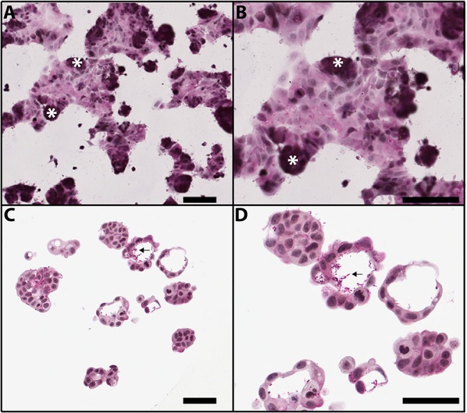
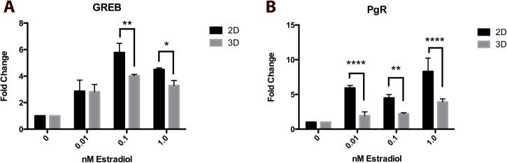
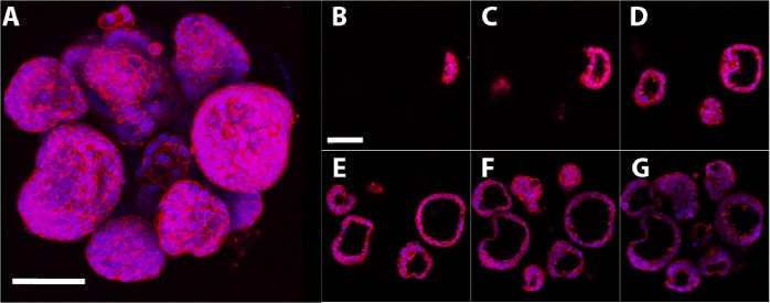
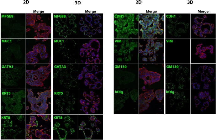
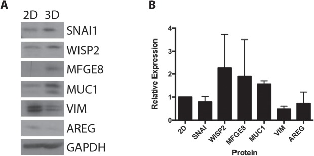
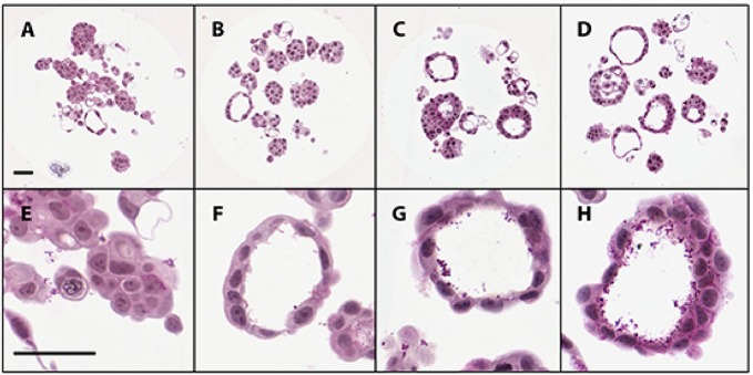
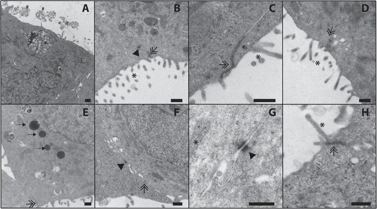
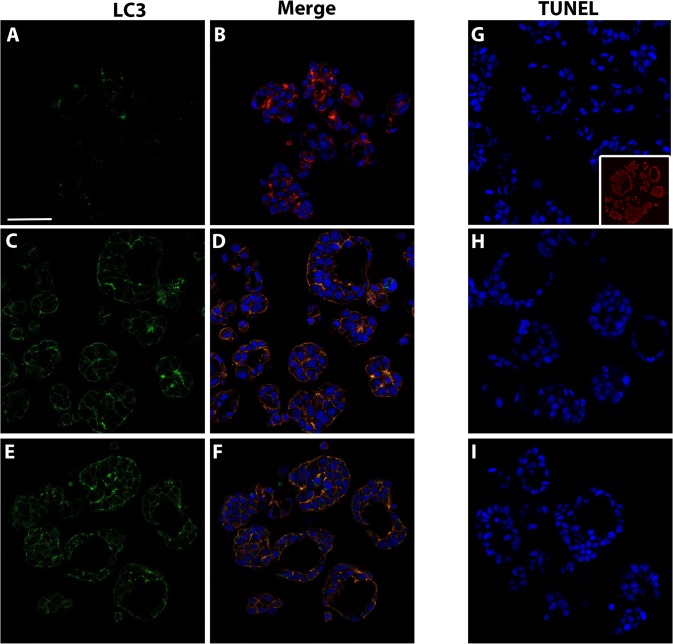
Three-dimensional (3D) cultures are increasing in use because of their ability to represent in vivo human physiology when compared to monolayer two-dimensional (2D) cultures. When grown in 3D using scaffold-free agarose hydrogels, MCF-7 human breast cancer cells self-organize to form directionally-oriented microtissues that contain a luminal space, reminiscent of the in vivo structure of the mammary gland. When compared to MCF-7 cells cultured in 2D monolayer culture, MCF-7 microtissues exhibit increased mRNA expression of luminal epithelial markers keratin 8 and keratin 19 and decreased expression of basal marker keratin 14 and the mesenchymal marker vimentin. These 3D MCF-7 microtissues remain responsive to estrogens, as demonstrated by induction of known estrogen target mRNAs following exposure to 17β-estradiol. Culture of MCF-7 cells in scaffold-free conditions allows for the formation of more differentiated, estrogen-responsive structures that are a more relevant system for evaluation of estrogenic compounds than traditional 2D models.
与单层二维(2D)培养相比,三维(3D)培养因其能够模拟体内人体生理状态而越来越多地被使用。当使用无支架琼脂糖水凝胶进行3D培养时,MCF-7人乳腺癌细胞会自我组织形成具有管腔空间的定向微组织,这让人联想到乳腺的体内结构。与在2D单层培养中培养的MCF-7细胞相比,MCF-7微组织中管腔上皮标志物角蛋白8和角蛋白19的mRNA表达增加,而基底标志物角蛋白14和间充质标志物波形蛋白的表达降低。这些3D MCF-7微组织对雌激素仍有反应,如在暴露于17β-雌二醇后已知雌激素靶mRNA的诱导所证明的那样。在无支架条件下培养MCF-7细胞能够形成更具分化性、对雌激素有反应的结构,与传统的2D模型相比,这是一个更适合评估雌激素化合物的系统。