Major Gretel, Ahn Minjun, Cho Won-Woo, Santos Miguel, Wise Jessika, Phillips Elisabeth, Wise Steven G, Jang Jinah, Rnjak-Kovacina Jelena, Woodfield Tim, Lim Khoon S
Department of Orthopaedic Surgery and Musculoskeletal Medicine, Centre for Bioengineering & Nanomedicine, University of Otago, Christchurch, New Zealand.
Pohang University of Science and Technology (POSTECH), Pohang, South Korea.
Mater Today Bio. 2024 Feb 16;25:101004. doi: 10.1016/j.mtbio.2024.101004. eCollection 2024 Apr.
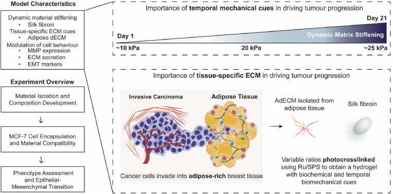
Extracellular matrix (ECM) stiffening is a common occurrence during the progression of many diseases, such as breast cancer. To accurately mimic the pathophysiological context of disease within 3D models, there is high demand for smart biomaterials which replicate the dynamic and temporal mechanical cues of diseased states. This study describes a preclinical disease model, using breast cancer as an example, which replicates the dynamic plasticity of the tumour microenvironment by incorporating temporal (3-week progression) biomechanical cues within a tissue-specific hydrogel microenvironment. The composite hydrogel formulation, integrating adipose-derived decellularised ECM (AdECM) and silk fibroin, was initially crosslinked using a visible light-mediated system, and then progressively stiffened through spontaneous secondary structure interactions inherent between the polymer chains (∼10-15 kPa increase, with a final stiffness of 25 kPa). When encapsulated and cultured , MCF-7 breast cancer cells initially formed numerous, large spheroids (>1000 μm in area), however, with progressive temporal stiffening, cells demonstrated growth arrest and underwent phenotypic changes resulting in intratumoral heterogeneity. Unlike widely-investigated static mechanical models, this stiffening hydrogel allowed for progressive phenotypic changes to be observed, and fostered the development of mature organoid-like spheroids, which mimicked both the organisation and acinar-structures of mature breast epithelium. The spheroids contained a central population of cells which expressed aggressive cellular programs, evidenced by increased fibronectin expression and reduction of E-cadherin. The phenotypic heterogeneity observed using this model is more reflective of physiological tumours, demonstrating the importance of establishing temporal cues within preclinical models in future work. Overall, the developed model demonstrated a novel strategy to uncouple ECM biomechanical properties from the cellular complexities of the disease microenvironment and offers the potential for wide applicability in other 3D disease models through addition of tissue-specific dECM materials.
细胞外基质(ECM)硬化在许多疾病(如乳腺癌)进展过程中普遍存在。为了在三维模型中准确模拟疾病的病理生理背景,对能够复制疾病状态动态和时间性力学线索的智能生物材料有很高的需求。本研究以乳腺癌为例描述了一种临床前疾病模型,该模型通过在组织特异性水凝胶微环境中纳入时间性(3周进展)生物力学线索来复制肿瘤微环境的动态可塑性。将脂肪来源的脱细胞ECM(AdECM)和丝素蛋白整合的复合水凝胶配方最初使用可见光介导系统进行交联,然后通过聚合物链之间固有的自发二级结构相互作用逐渐硬化(增加约10 - 15kPa,最终硬度为25kPa)。当封装并培养时,MCF - 7乳腺癌细胞最初形成许多大球体(面积>1000μm),然而,随着时间的逐渐硬化,细胞表现出生长停滞并发生表型变化,导致肿瘤内异质性。与广泛研究的静态力学模型不同,这种硬化水凝胶允许观察到渐进的表型变化,并促进了成熟类器官样球体的形成,其模仿了成熟乳腺上皮的组织结构和腺泡结构。球体包含一群表达侵袭性细胞程序的中央细胞,这通过纤连蛋白表达增加和E - 钙黏蛋白减少得到证明。使用该模型观察到的表型异质性更能反映生理性肿瘤,表明在未来工作中在临床前模型中建立时间线索的重要性。总体而言,所开发的模型展示了一种将ECM生物力学特性与疾病微环境的细胞复杂性解耦的新策略,并通过添加组织特异性dECM材料为其他三维疾病模型的广泛应用提供了潜力。