Herling Lotta, Johnson Jonas, Ferm-Widlund Kjerstin, Lindgren Peter, Acharya Ganesh, Westgren Magnus
Centre for Fetal Medicine, Department of Obstetrics and Gynecology, Karolinska University Hospital, Stockholm, Sweden.
University Hospital of Northern Norway, Tromsø, Norway.
Cardiovasc Ultrasound. 2015 Aug 27;13:39. doi: 10.1186/s12947-015-0034-3.
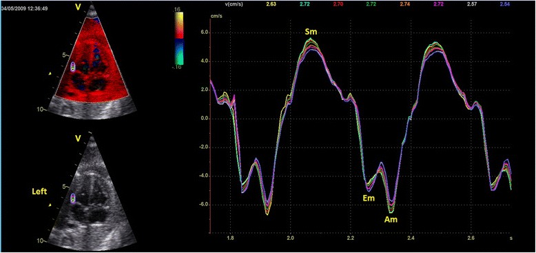
Tissue Doppler imaging (TDI) can be used to assess fetal cardiac function and it has been shown to detect changes associated with hypoxia in animal models. However, the analysis is cumbersome and time consuming. The main objective of this study was to evaluate the feasibility of a new algorithm developed for the automated analysis of color TDI velocity recordings of the fetal myocardium. Furthermore, we wanted to assess the effect of different sizes of region of interests (ROI) on the measurement of cardiac cycle time intervals and myocardial velocities at different gestations.
This study included analysis of 261 TDI velocity traces obtained from 17 fetal echocardiographic examinations performed longitudinally on five pregnant women. Cine-loops of fetal cardiac four chamber view were recorded with color overlay in TDI mode and stored for off-line analysis. ROIs of different sizes were placed at the level of the atrioventricular plane in the septum and in the right and left ventricular walls of the fetal heart. An automated algorithm was then used for the analysis of velocity traces.
Out of the total 261 velocity traces, it was possible to analyze 203 (78 %) traces with the automated algorithm. It was possible to analyze 93 % (81/87) of traces recorded from the right ventricular wall, 82 % (71/87) from the left ventricular wall and 59 % (51/87) from the septum. There was a trend towards decreasing myocardial velocities with increasing ROI length. However, the cardiac cycle time intervals were similar irrespective of which ROI size was used.
An automated analysis of color TDI fetal myocardial velocity traces seems feasible, especially for measuring cardiac cycle time intervals, and has the potential for clinical application.
组织多普勒成像(TDI)可用于评估胎儿心脏功能,并且已证实在动物模型中它能检测到与缺氧相关的变化。然而,分析过程繁琐且耗时。本研究的主要目的是评估一种新算法用于自动分析胎儿心肌彩色TDI速度记录的可行性。此外,我们想评估不同大小的感兴趣区域(ROI)对不同孕周心脏周期时间间隔和心肌速度测量的影响。
本研究包括对从5名孕妇纵向进行的17次胎儿超声心动图检查中获得的261条TDI速度轨迹进行分析。以TDI模式记录胎儿心脏四腔心切面的电影环,并叠加彩色,存储用于离线分析。在胎儿心脏的间隔以及右心室壁和左心室壁的房室平面水平放置不同大小的ROI。然后使用一种自动算法分析速度轨迹。
在总共261条速度轨迹中,使用自动算法能够分析203条(78%)轨迹。能够分析从右心室壁记录的93%(81/87)的轨迹、从左心室壁记录的82%(71/87)的轨迹以及从间隔记录的59%(51/87)的轨迹。随着ROI长度增加,心肌速度有下降趋势。然而,无论使用哪种ROI大小,心脏周期时间间隔相似。
对胎儿心肌彩色TDI速度轨迹进行自动分析似乎是可行的,特别是用于测量心脏周期时间间隔,并且具有临床应用潜力。