Menegas William, Bergan Joseph F, Ogawa Sachie K, Isogai Yoh, Umadevi Venkataraju Kannan, Osten Pavel, Uchida Naoshige, Watabe-Uchida Mitsuko
Center for Brain Science, Department of Molecular and Cellular Biology, Harvard University, Cambridge, United States.
Cold Spring Harbor Laboratory, Cold Spring Harbor, United States.
Elife. 2015 Aug 31;4:e10032. doi: 10.7554/eLife.10032.
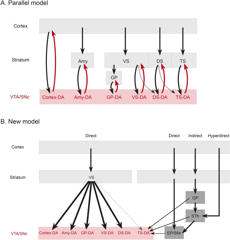
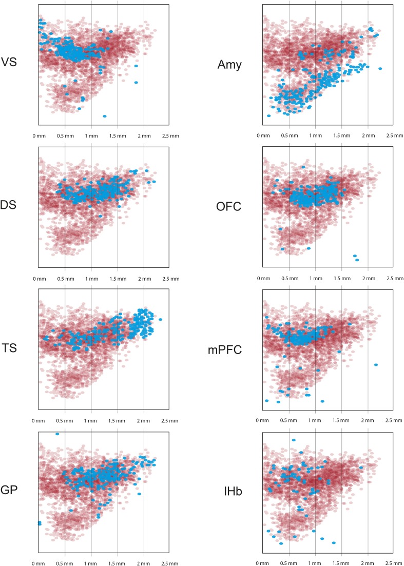
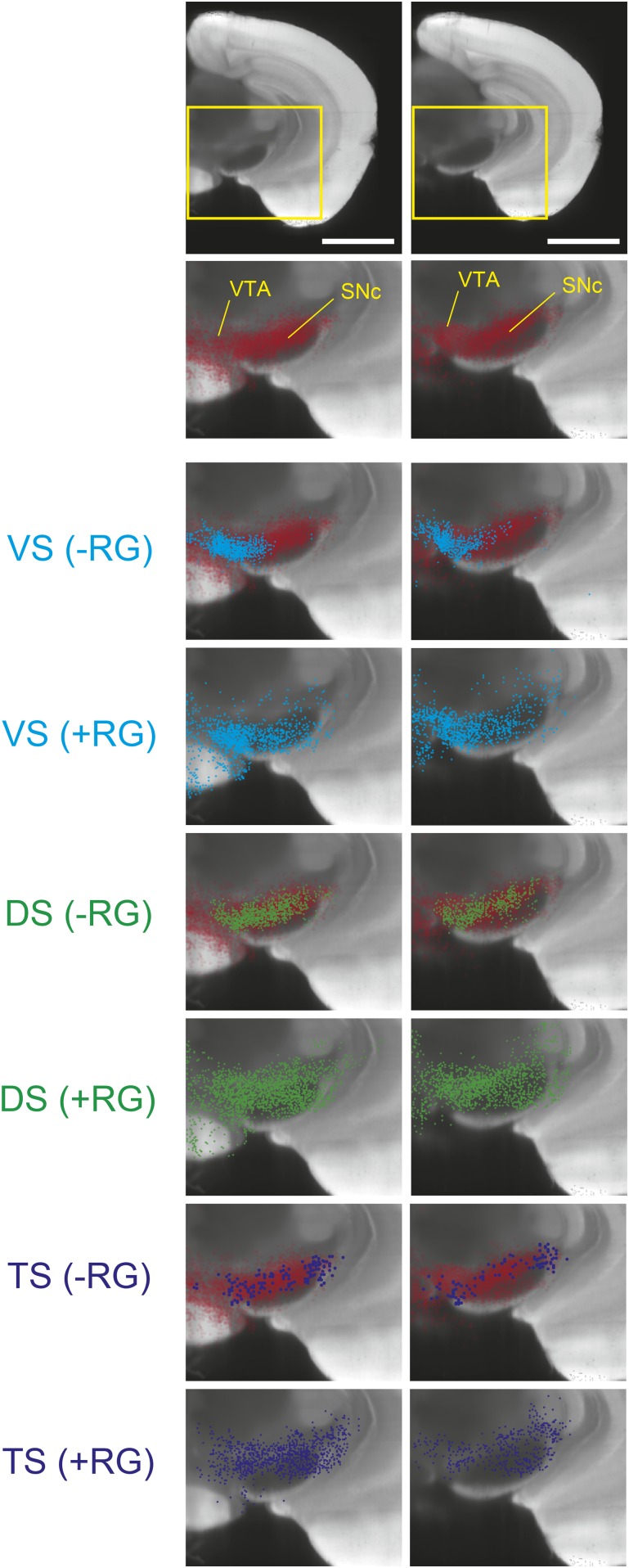
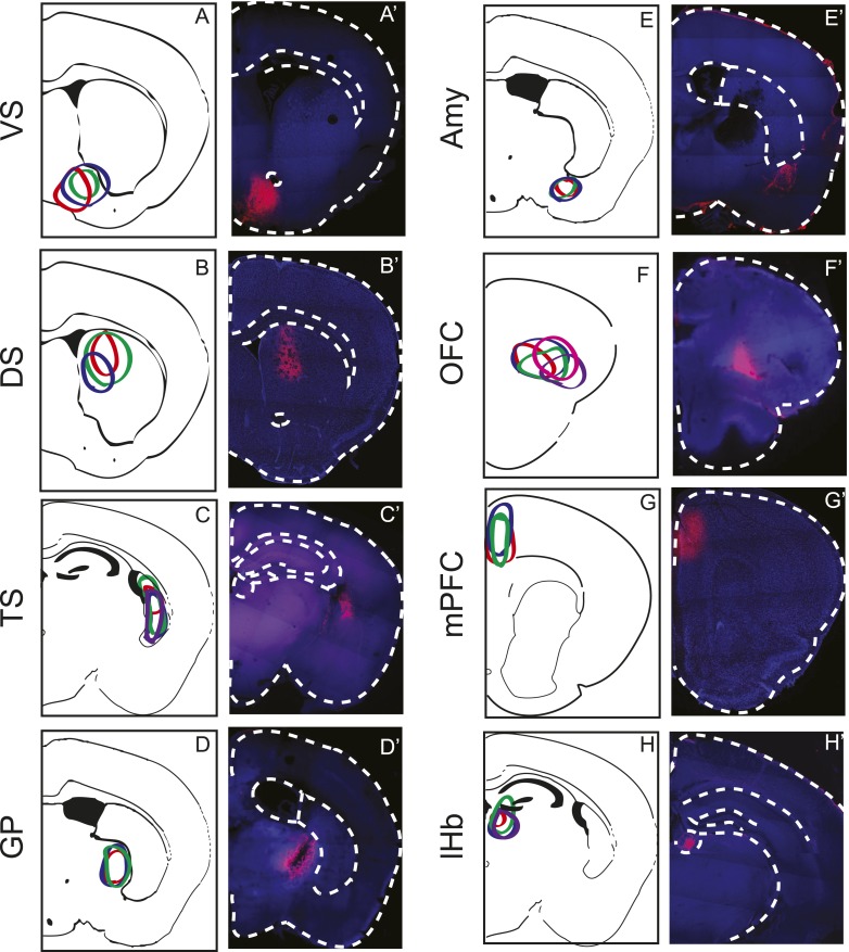
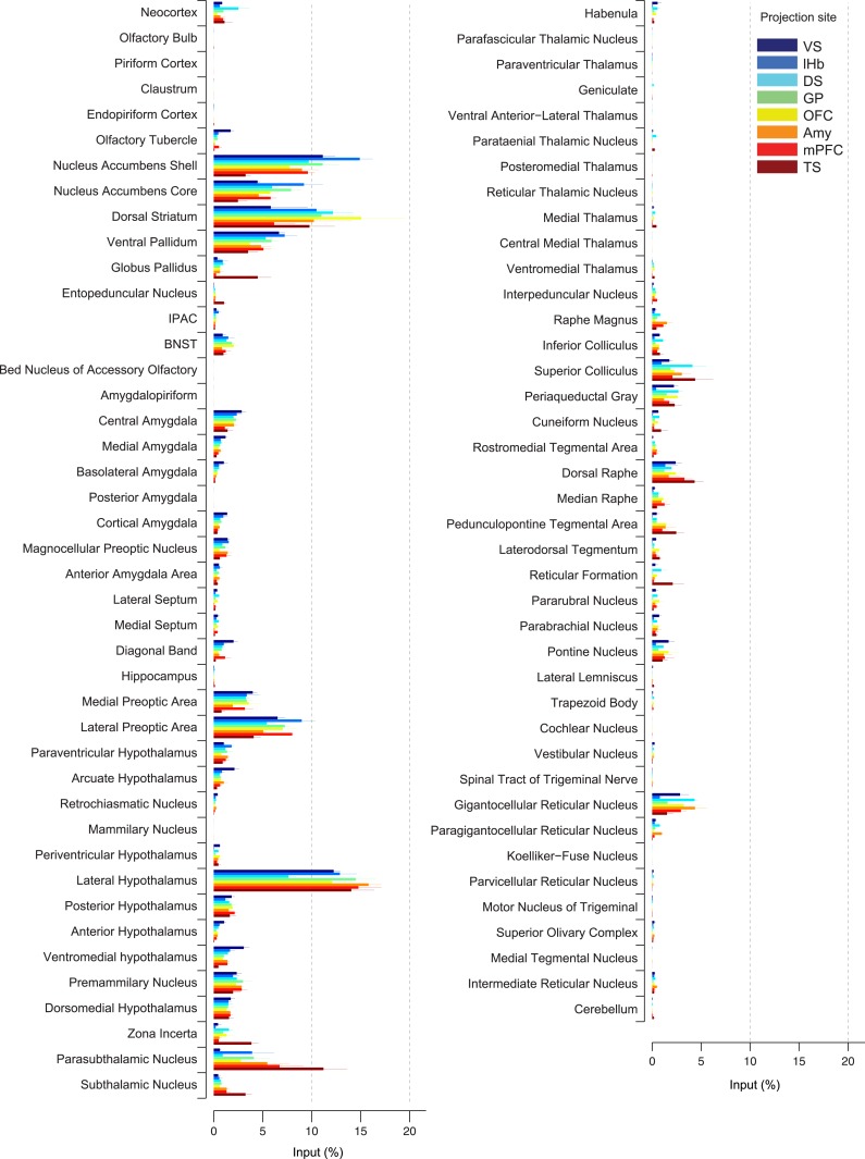
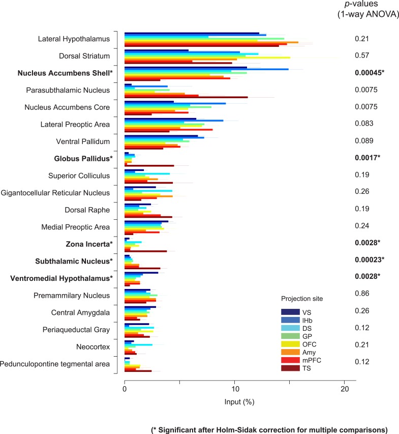
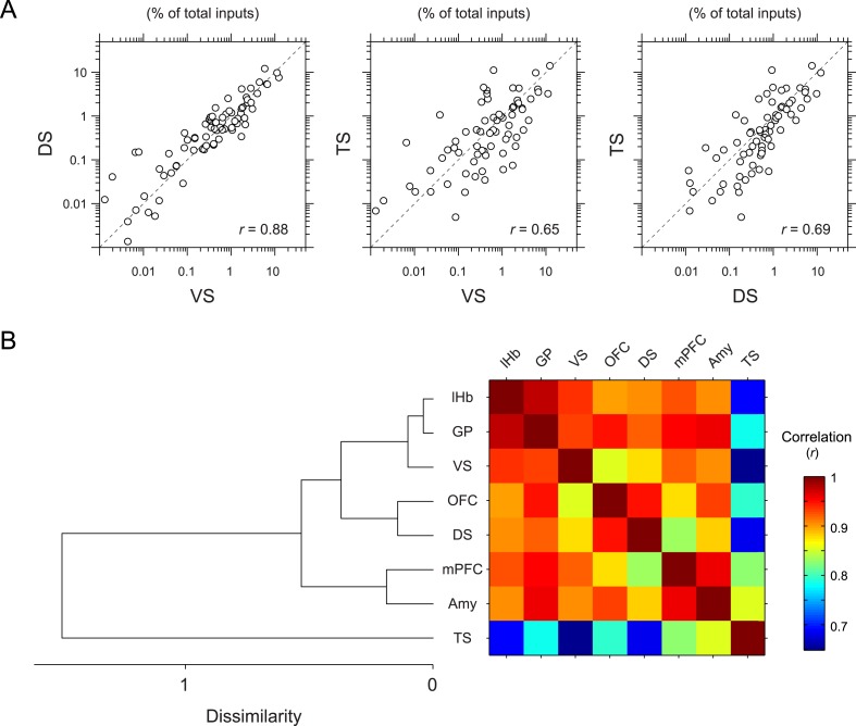
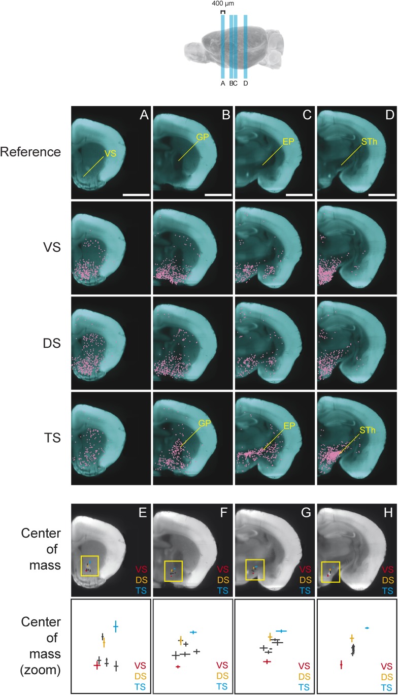
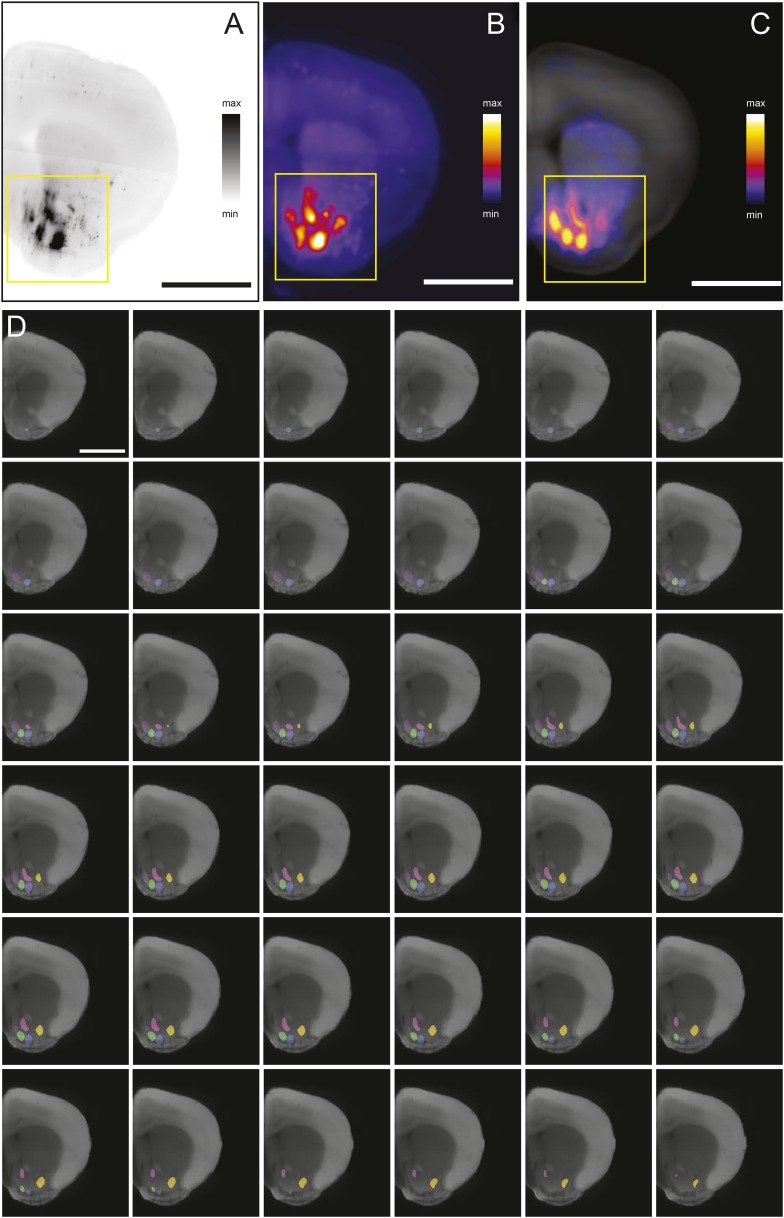
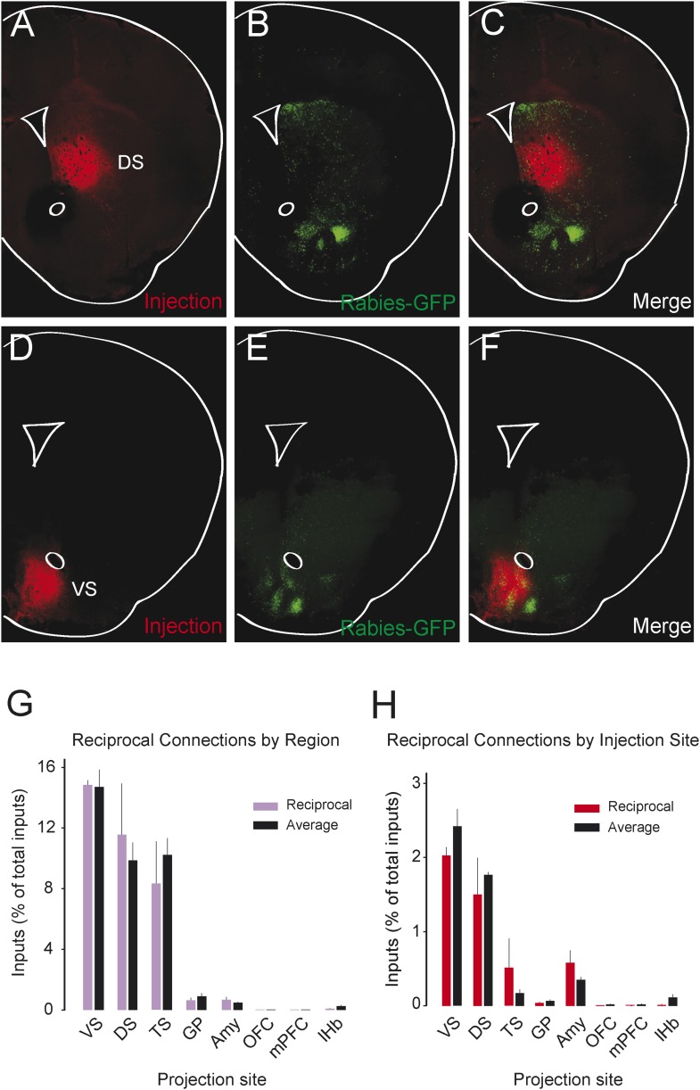
Combining rabies-virus tracing, optical clearing (CLARITY), and whole-brain light-sheet imaging, we mapped the monosynaptic inputs to midbrain dopamine neurons projecting to different targets (different parts of the striatum, cortex, amygdala, etc) in mice. We found that most populations of dopamine neurons receive a similar set of inputs rather than forming strong reciprocal connections with their target areas. A common feature among most populations of dopamine neurons was the existence of dense 'clusters' of inputs within the ventral striatum. However, we found that dopamine neurons projecting to the posterior striatum were outliers, receiving relatively few inputs from the ventral striatum and instead receiving more inputs from the globus pallidus, subthalamic nucleus, and zona incerta. These results lay a foundation for understanding the input/output structure of the midbrain dopamine circuit and demonstrate that dopamine neurons projecting to the posterior striatum constitute a unique class of dopamine neurons regulated by different inputs.
结合狂犬病病毒示踪、光学清透技术(CLARITY)和全脑光片成像技术,我们绘制了小鼠中脑多巴胺能神经元投射到不同靶点(纹状体、皮质、杏仁核等不同部位)的单突触输入图谱。我们发现,大多数多巴胺能神经元群体接收的输入集相似,而非与其靶区域形成强相互连接。大多数多巴胺能神经元群体的一个共同特征是腹侧纹状体内存在密集的输入“簇”。然而,我们发现投射到纹状体后部的多巴胺能神经元是个例外,它们从腹侧纹状体接收的输入相对较少,反而从苍白球、丘脑底核和未定带接收更多输入。这些结果为理解中脑多巴胺回路的输入/输出结构奠定了基础,并表明投射到纹状体后部的多巴胺能神经元构成了一类受不同输入调控的独特多巴胺能神经元。