• 文献检索
  • 文档翻译
  • 深度研究
  • 学术资讯
  • Suppr Zotero 插件Zotero 插件
  • 邀请有礼
  • 套餐&价格
  • 历史记录
应用&插件
Suppr Zotero 插件Zotero 插件浏览器插件Mac 客户端Windows 客户端微信小程序
定价
高级版会员购买积分包购买API积分包
服务
文献检索文档翻译深度研究API 文档MCP 服务
关于我们
关于 Suppr公司介绍联系我们用户协议隐私条款
关注我们

Suppr 超能文献

核心技术专利:CN118964589B侵权必究
粤ICP备2023148730 号-1Suppr @ 2026

文献检索

告别复杂PubMed语法,用中文像聊天一样搜索,搜遍4000万医学文献。AI智能推荐,让科研检索更轻松。

立即免费搜索

文件翻译

保留排版,准确专业,支持PDF/Word/PPT等文件格式,支持 12+语言互译。

免费翻译文档

深度研究

AI帮你快速写综述,25分钟生成高质量综述,智能提取关键信息,辅助科研写作。

立即免费体验

骨保护素在软骨细胞中的特异性表达调节干骺端骨中的破骨细胞形成。

Chondrocytes-Specific Expression of Osteoprotegerin Modulates Osteoclast Formation in Metaphyseal Bone.

作者信息

Wang Baoli, Jin Hongting, Shu Bing, Mira Ranim R, Chen Di

机构信息

Key Lab of Hormone and Development (Ministry of Health), Metabolic Diseases Hospital and Tianjin Institute of Endocrinology, Tianjin Medical University, Tianjin 300070, China.

Department of Orthopaedics, Center for Musculoskeletal Research, University of Rochester School of Medicine, Rochester, NY 14642, USA.

出版信息

Sci Rep. 2015 Sep 2;5:13667. doi: 10.1038/srep13667.

DOI:10.1038/srep13667
PMID:26329493
原文链接:https://pmc.ncbi.nlm.nih.gov/articles/PMC4556963/
Abstract

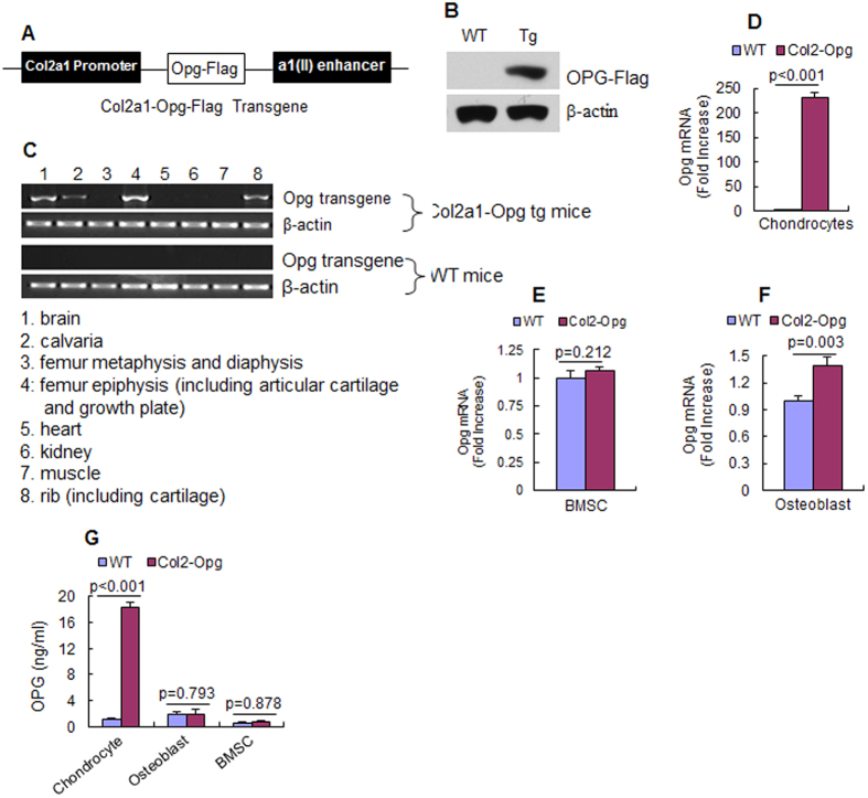
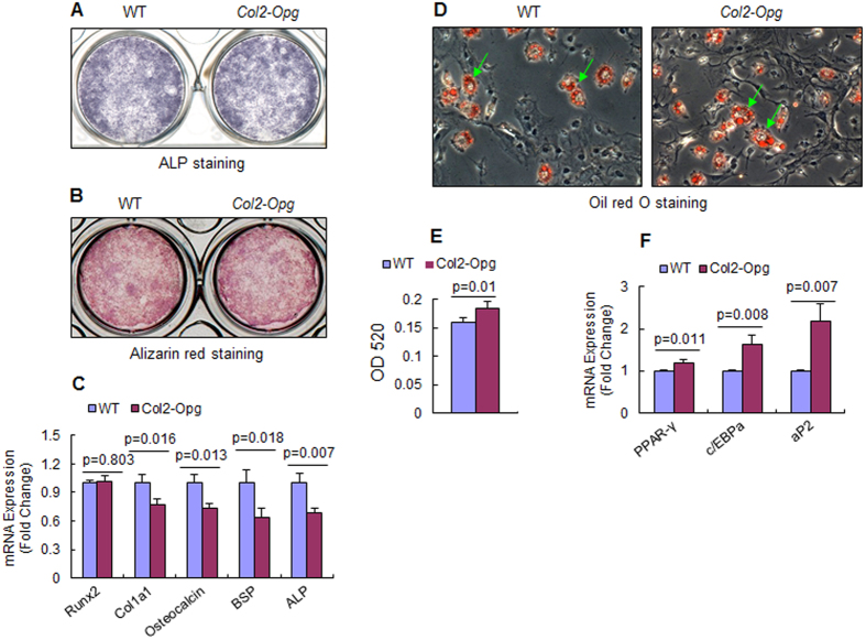
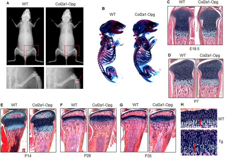
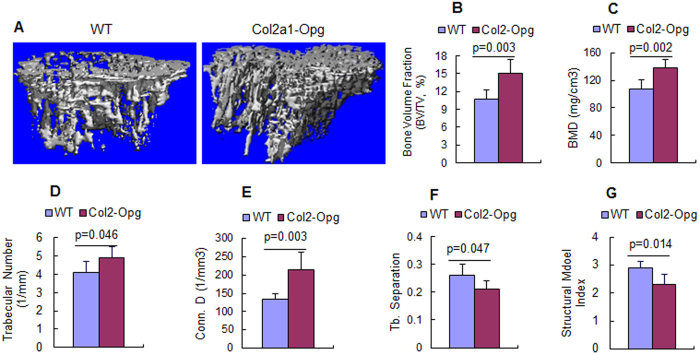
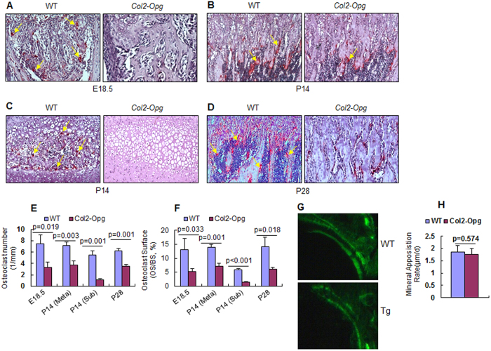
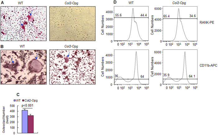
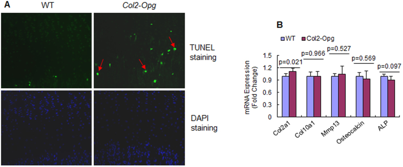
Bone marrow stromal cells/osteoblasts were originally thought to be the major player in regulating osteoclast differentiation through expressing RANKL/OPG cytokines. Recent studies have established that chondrocytes also express RANKL/OPG and support osteoclast formation. Till now, the in vivo function of chondrocyte-produced OPG in osteoclast formation and postnatal bone growth has not been directly investigated. In this study, chondrocyte-specific Opg transgenic mice were generated by using type II collagen promoter. The Col2-Opg transgenic mice showed delayed formation of secondary ossification center and localized increase of bone mass in proximal metaphysis of tibiae. TRAP staining showed that osteoclast numbers were reduced in both secondary ossification center and proximal metaphysis. This finding was further confirmed by in vitro chondrocyte/spleen cell co-culture assay. In contrast, the mineral apposition rates were not changed in Col2-Opg transgenic mice. TUNEL staining revealed more apoptotic hypertrophic chondrocytes in the growth plate of Col2-Opg mice. Flow cytometry analysis showed fewer RANK-expressing cells in the marrow of Col2a1-Opg mice, suggesting the role of OPG in blocking the differentiation of early mesenchymal progenitors into RANK-expressing pre-osteoclasts. Our results demonstrated that OPG expression in chondrocyte increases bone mass in the proximal metaphysis of tibiae through negative regulation of osteoclast formation.

摘要

骨髓基质细胞/成骨细胞最初被认为是通过表达RANKL/OPG细胞因子来调节破骨细胞分化的主要细胞。最近的研究表明,软骨细胞也表达RANKL/OPG并支持破骨细胞形成。到目前为止,软骨细胞产生的OPG在破骨细胞形成和出生后骨骼生长中的体内功能尚未得到直接研究。在本研究中,通过使用II型胶原启动子生成了软骨细胞特异性Opg转基因小鼠。Col2-Opg转基因小鼠显示出二级骨化中心形成延迟,胫骨近端干骺端骨量局部增加。TRAP染色显示,二级骨化中心和近端干骺端的破骨细胞数量均减少。体外软骨细胞/脾细胞共培养试验进一步证实了这一发现。相比之下,Col2-Opg转基因小鼠的矿物质沉积率没有变化。TUNEL染色显示Col2-Opg小鼠生长板中凋亡的肥大软骨细胞更多。流式细胞术分析显示Col2a1-Opg小鼠骨髓中表达RANK的细胞较少,提示OPG在阻止早期间充质祖细胞分化为表达RANK的破骨细胞前体细胞中的作用。我们的结果表明,软骨细胞中OPG的表达通过对破骨细胞形成的负调节增加了胫骨近端干骺端的骨量。

https://cdn.ncbi.nlm.nih.gov/pmc/blobs/2046/4556963/3c01c2e569b4/srep13667-f7.jpg
https://cdn.ncbi.nlm.nih.gov/pmc/blobs/2046/4556963/af2ed692ada7/srep13667-f1.jpg
https://cdn.ncbi.nlm.nih.gov/pmc/blobs/2046/4556963/37170051ce5e/srep13667-f2.jpg
https://cdn.ncbi.nlm.nih.gov/pmc/blobs/2046/4556963/e7189a641dc9/srep13667-f3.jpg
https://cdn.ncbi.nlm.nih.gov/pmc/blobs/2046/4556963/558e03736fe3/srep13667-f4.jpg
https://cdn.ncbi.nlm.nih.gov/pmc/blobs/2046/4556963/32b64c791416/srep13667-f5.jpg
https://cdn.ncbi.nlm.nih.gov/pmc/blobs/2046/4556963/f269909226d2/srep13667-f6.jpg
https://cdn.ncbi.nlm.nih.gov/pmc/blobs/2046/4556963/3c01c2e569b4/srep13667-f7.jpg

相似文献

1
Chondrocytes-Specific Expression of Osteoprotegerin Modulates Osteoclast Formation in Metaphyseal Bone.骨保护素在软骨细胞中的特异性表达调节干骺端骨中的破骨细胞形成。
Sci Rep. 2015 Sep 2;5:13667. doi: 10.1038/srep13667.
2
Effects of osteoprotegerin administration on osteoclast differentiation and trabecular bone structure in osteoprotegerin-deficient mice.给予骨保护素对骨保护素缺乏小鼠破骨细胞分化及小梁骨结构的影响。
J Electron Microsc (Tokyo). 2005 Oct;54(5):467-77. doi: 10.1093/jmicro/dfi066. Epub 2005 Dec 9.
3
Aging increases stromal/osteoblastic cell-induced osteoclastogenesis and alters the osteoclast precursor pool in the mouse.衰老会增加基质/成骨细胞诱导的破骨细胞生成,并改变小鼠体内破骨细胞前体细胞库。
J Bone Miner Res. 2005 Sep;20(9):1659-68. doi: 10.1359/JBMR.050503. Epub 2005 May 2.
4
Chondrocyte FGFR3 Regulates Bone Mass by Inhibiting Osteogenesis.软骨细胞FGFR3通过抑制成骨作用调节骨量。
J Biol Chem. 2016 Nov 25;291(48):24912-24921. doi: 10.1074/jbc.M116.730093. Epub 2016 Oct 11.
5
Deletion of beta catenin in hypertrophic growth plate chondrocytes impairs trabecular bone formation.β-连环蛋白在肥大的生长板软骨细胞中的缺失会损害小梁骨的形成。
Bone. 2013 Jul;55(1):102-12. doi: 10.1016/j.bone.2013.03.019. Epub 2013 Apr 6.
6
Chondrocyte β-catenin signaling regulates postnatal bone remodeling through modulation of osteoclast formation in a murine model.软骨细胞β-catenin 信号通过调节破骨细胞形成调节小鼠模型中的出生后骨重塑。
Arthritis Rheumatol. 2014 Jan;66(1):107-20. doi: 10.1002/art.38195.
7
The cross-talk between osteoclasts and osteoblasts in response to strontium treatment: involvement of osteoprotegerin.成骨细胞与破骨细胞间的串扰在锶治疗中的反应:骨保护素的参与。
Bone. 2011 Dec;49(6):1290-8. doi: 10.1016/j.bone.2011.08.031. Epub 2011 Sep 9.
8
Murine and chicken chondrocytes regulate osteoclastogenesis by producing RANKL in response to BMP2.小鼠和鸡软骨细胞通过响应骨形态发生蛋白2(BMP2)产生核因子κB受体活化因子配体(RANKL)来调节破骨细胞生成。
J Bone Miner Res. 2008 Mar;23(3):314-25. doi: 10.1359/jbmr.071025.
9
Osteoprotegerin produced by osteoblasts is an important regulator in osteoclast development and function.成骨细胞产生的骨保护素是破骨细胞发育和功能的重要调节因子。
Endocrinology. 2000 Sep;141(9):3478-84. doi: 10.1210/endo.141.9.7634.
10
Production of IL-7 is increased in ovariectomized mice, but not RANKL mRNA expression by osteoblasts/stromal cells in bone, and IL-7 enhances generation of osteoclast precursors in vitro.在去卵巢小鼠中,IL-7的产生增加,但骨中破骨细胞分化因子(RANKL)的mRNA表达未受影响,且IL-7在体外可增强破骨细胞前体的生成。
J Bone Miner Metab. 2007;25(1):19-27. doi: 10.1007/s00774-006-0723-y. Epub 2007 Jan 1.

引用本文的文献

1
Biomaterial Cues for Regulation of Osteoclast Differentiation and Function in Bone Regeneration.用于调节骨再生中破骨细胞分化和功能的生物材料线索
Adv Ther (Weinh). 2025 Jan;8(1). doi: 10.1002/adtp.202400296. Epub 2024 Nov 15.
2
The Pathogenetic Role of RANK/RANKL/OPG Signaling in Osteoarthritis and Related Targeted Therapies.RANK/RANKL/OPG信号通路在骨关节炎中的致病作用及相关靶向治疗
Biomedicines. 2024 Oct 10;12(10):2292. doi: 10.3390/biomedicines12102292.
3
Proton and Carbon Ion Irradiation Changes the Process of Endochondral Ossification in an Ex Vivo Femur Organotypic Culture Model.

本文引用的文献

1
Chondrocytes transdifferentiate into osteoblasts in endochondral bone during development, postnatal growth and fracture healing in mice.在小鼠的发育、出生后生长和骨折愈合过程中,软骨细胞在软骨内骨中会转分化为成骨细胞。
PLoS Genet. 2014 Dec 4;10(12):e1004820. doi: 10.1371/journal.pgen.1004820. eCollection 2014 Dec.
2
A subset of chondrogenic cells provides early mesenchymal progenitors in growing bones.在生长中的骨骼中,软骨细胞亚群为早期间充质祖细胞提供来源。
Nat Cell Biol. 2014 Dec;16(12):1157-67. doi: 10.1038/ncb3067. Epub 2014 Nov 24.
3
Col2CreER(T2), a mouse model for a chondrocyte-specific and inducible gene deletion.
质子和碳离子辐照改变体外股骨器官培养模型的软骨内骨化过程。
Cells. 2023 Sep 18;12(18):2301. doi: 10.3390/cells12182301.
4
The role of cells and signal pathways in subchondral bone in osteoarthritis.细胞和信号通路在骨关节炎软骨下骨中的作用。
Bone Joint Res. 2023 Sep 8;12(9):536-545. doi: 10.1302/2046-3758.129.BJR-2023-0081.R1.
5
Exercise improves subchondral bone microenvironment through regulating bone-cartilage crosstalk.运动通过调节骨-软骨串扰改善软骨下骨微环境。
Front Endocrinol (Lausanne). 2023 May 23;14:1159393. doi: 10.3389/fendo.2023.1159393. eCollection 2023.
6
Biochemical characterization of a disease-causing human osteoprotegerin variant.一种导致疾病的人类护骨素变体的生化特征。
Sci Rep. 2022 Sep 10;12(1):15279. doi: 10.1038/s41598-022-19522-9.
7
Ablation of the miRNA cluster 24 in cartilage and osteoblasts impairs bone remodeling.软骨和成骨细胞中 miRNA 簇 24 的消融会损害骨重塑。
Sci Rep. 2022 Jun 1;12(1):9116. doi: 10.1038/s41598-022-13231-z.
8
IκB-ζ signaling promotes chondrocyte inflammatory phenotype, senescence, and erosive joint pathology.IκB-ζ信号传导促进软骨细胞炎症表型、衰老和侵蚀性关节病变。
Bone Res. 2022 Feb 11;10(1):12. doi: 10.1038/s41413-021-00183-9.
9
Subchondral bone microenvironment in osteoarthritis and pain.骨关节炎与疼痛中的软骨下骨微环境
Bone Res. 2021 Mar 17;9(1):20. doi: 10.1038/s41413-021-00147-z.
10
MINDIN Exerts Protumorigenic Actions on Primary Prostate Tumors via Downregulation of the Scaffold Protein NHERF-1.MINDIN通过下调支架蛋白NHERF-1对原发性前列腺肿瘤发挥促肿瘤作用。
Cancers (Basel). 2021 Jan 24;13(3):436. doi: 10.3390/cancers13030436.
Col2CreER(T2),一种用于软骨细胞特异性和诱导性基因缺失的小鼠模型。
Eur Cell Mater. 2014 Oct 23;28:236-45. doi: 10.22203/ecm.v028a16.
4
Hypertrophic chondrocytes can become osteoblasts and osteocytes in endochondral bone formation.肥大的软骨细胞可在软骨内骨形成中成为成骨细胞和骨细胞。
Proc Natl Acad Sci U S A. 2014 Aug 19;111(33):12097-102. doi: 10.1073/pnas.1302703111. Epub 2014 Aug 4.
5
Chondrocyte β-catenin signaling regulates postnatal bone remodeling through modulation of osteoclast formation in a murine model.软骨细胞β-catenin 信号通过调节破骨细胞形成调节小鼠模型中的出生后骨重塑。
Arthritis Rheumatol. 2014 Jan;66(1):107-20. doi: 10.1002/art.38195.
6
Zoledronic acid inhibits RANK expression and migration of osteoclast precursors during osteoclastogenesis.唑来膦酸在破骨细胞发生过程中抑制破骨细胞前体的 RANK 表达和迁移。
Naunyn Schmiedebergs Arch Pharmacol. 2011 Mar;383(3):297-308. doi: 10.1007/s00210-010-0596-4. Epub 2011 Jan 12.
7
Alcian blue/alizarin red staining of cartilage and bone in mouse.小鼠软骨和骨的阿尔新蓝/茜素红染色
Cold Spring Harb Protoc. 2009 Mar;2009(3):pdb.prot5170. doi: 10.1101/pdb.prot5170.
8
Axin2 controls bone remodeling through the beta-catenin-BMP signaling pathway in adult mice.在成年小鼠中,Axin2通过β-连环蛋白-骨形态发生蛋白信号通路控制骨重塑。
J Cell Sci. 2009 Oct 1;122(Pt 19):3566-78. doi: 10.1242/jcs.051904. Epub 2009 Sep 8.
9
Growth plate mechanics and mechanobiology. A survey of present understanding.生长板力学与机械生物学。当前认识概述。
J Biomech. 2009 Aug 25;42(12):1793-803. doi: 10.1016/j.jbiomech.2009.05.021. Epub 2009 Jun 21.
10
Inhibition of beta-catenin signaling causes defects in postnatal cartilage development.β-连环蛋白信号通路的抑制会导致出生后软骨发育缺陷。
J Cell Sci. 2008 May 1;121(Pt 9):1455-65. doi: 10.1242/jcs.020362. Epub 2008 Apr 8.