Juras Vladimir, Bohndorf Klaus, Heule Rahel, Kronnerwetter Claudia, Szomolanyi Pavol, Hager Benedikt, Bieri Oliver, Zbyn Stefan, Trattnig Siegfried
High Field MR Centre, Department of Biomedical Imaging and Image-Guided Therapy, Medical University of Vienna, Vienna, Austria.
Department of Imaging Methods, Institute of Measurement Science, Bratislava, Slovakia.
Eur Radiol. 2016 Jun;26(6):1905-12. doi: 10.1007/s00330-015-3979-6. Epub 2015 Sep 3.
To assess the clinical relevance of T2 relaxation times, measured by 3D triple-echo steady-state (3D-TESS), in knee articular cartilage compared to conventional multi-echo spin-echo T2-mapping.
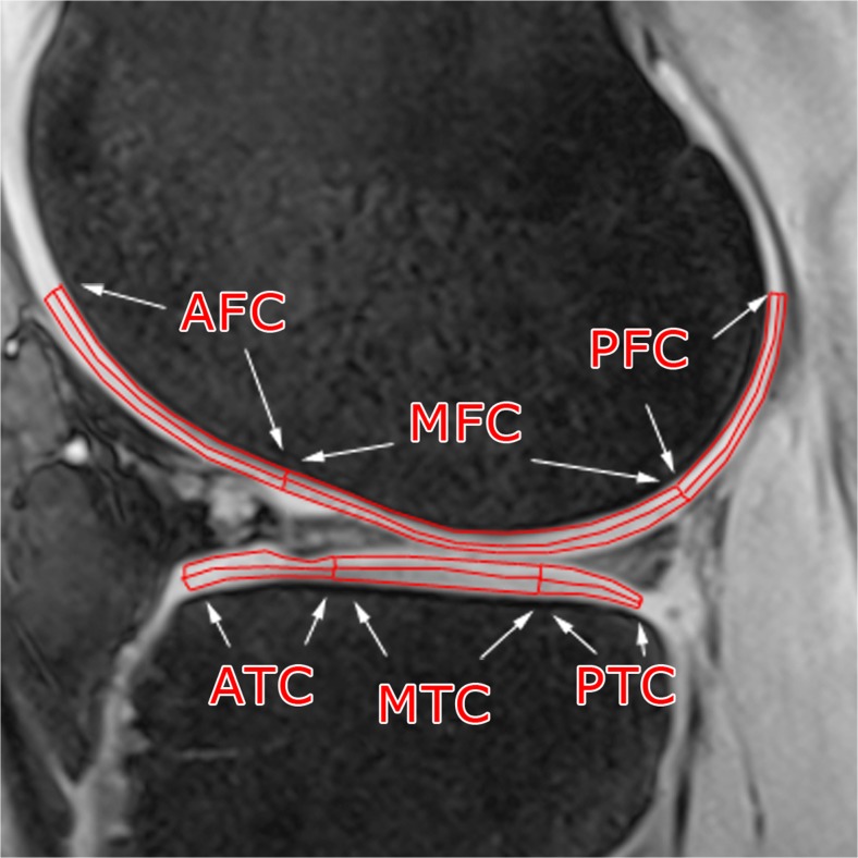
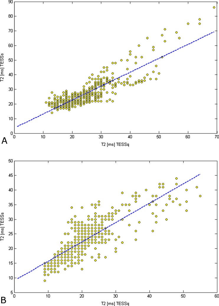
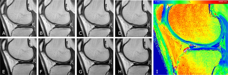
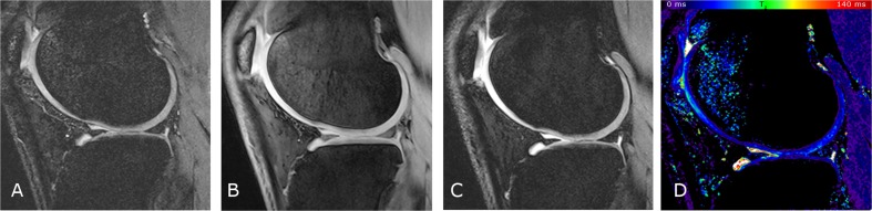
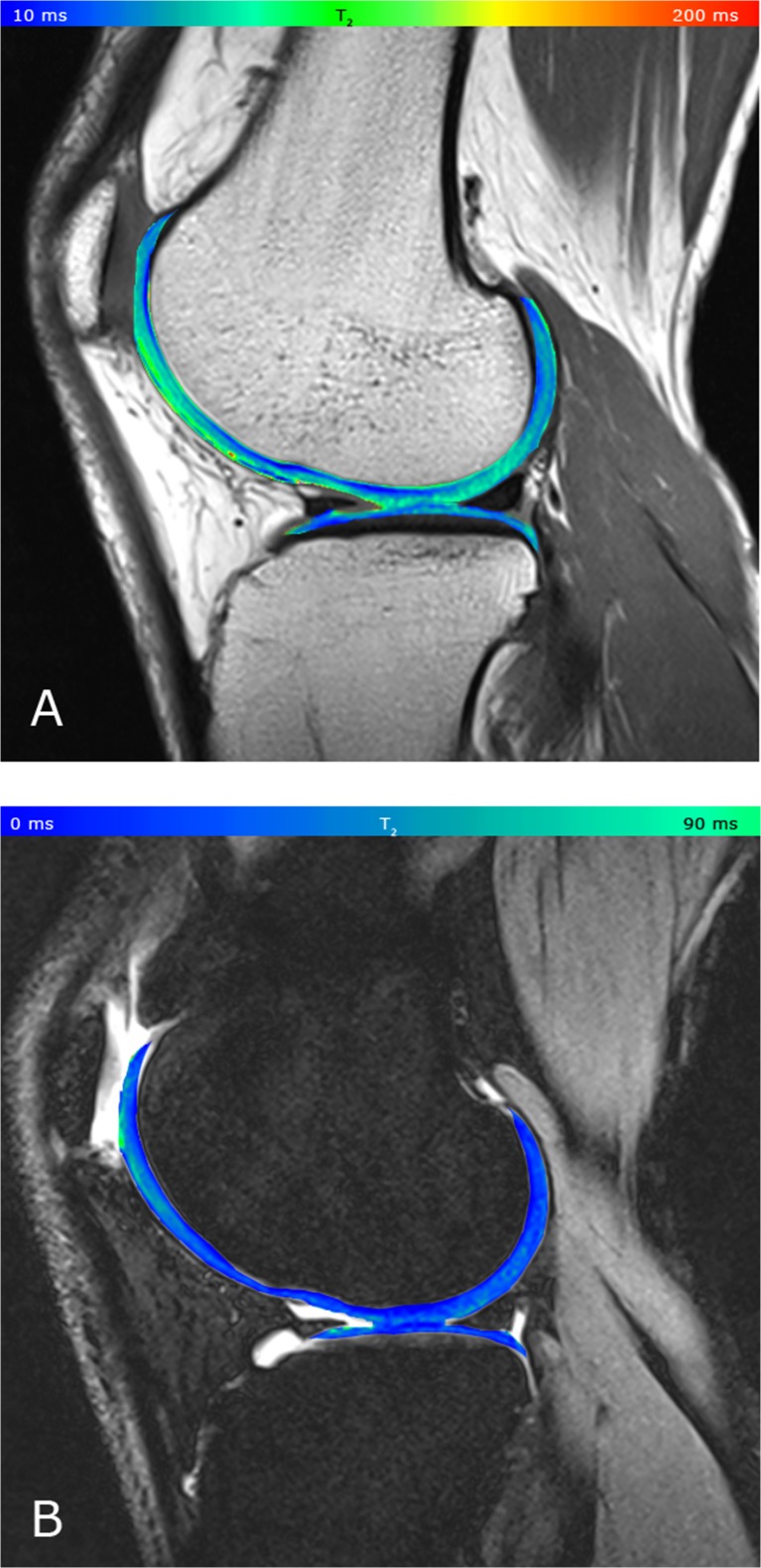
Thirteen volunteers and ten patients with focal cartilage lesions were included in this prospective study. All subjects underwent 3-Tesla MRI consisting of a multi-echo multi-slice spin-echo sequence (CPMG) as a reference method for T2 mapping, and 3D TESS with the same geometry settings, but variable acquisition times: standard (TESSs 4:35min) and quick (TESSq 2:05min). T2 values were compared in six different regions in the femoral and tibial cartilage using a Wilcoxon signed ranks test and the Pearson correlation coefficient (r). The local ethics committee approved this study, and all participants gave written informed consent.
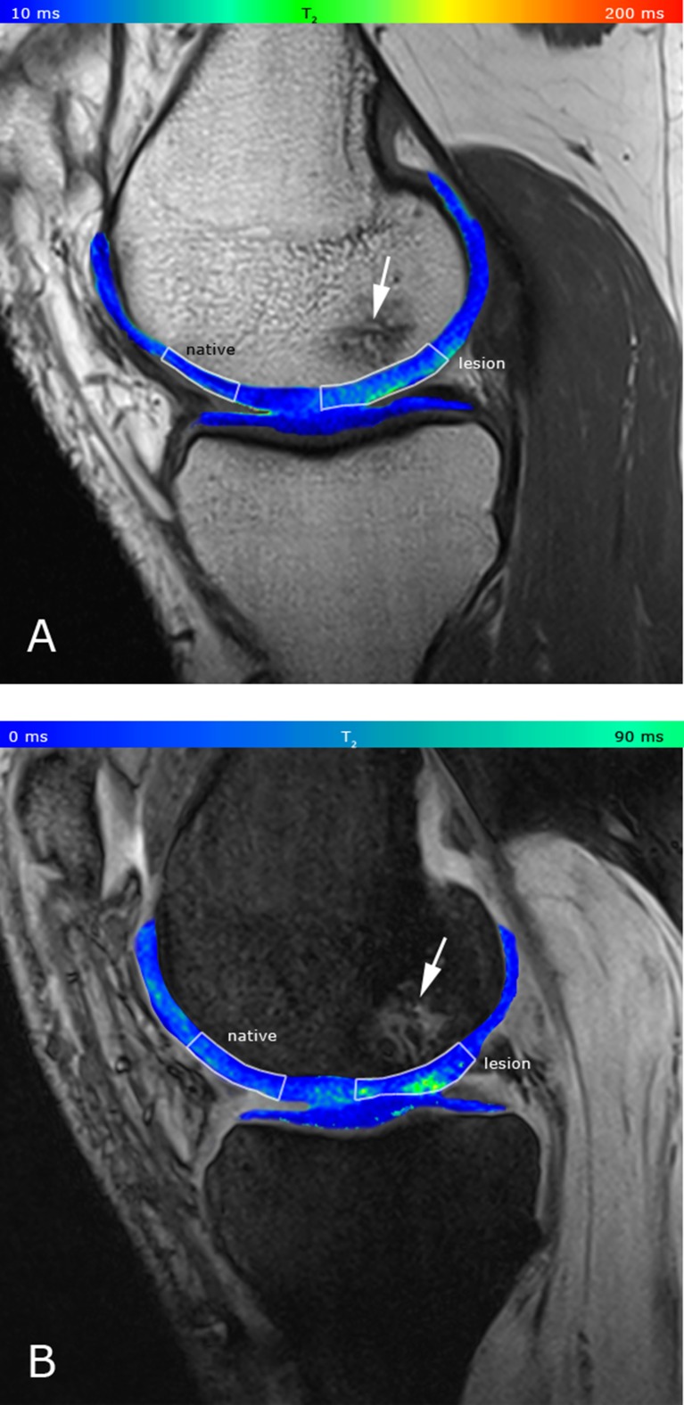
The mean quantitative T2 values measured by CPMG (mean: 46±9ms) in volunteers were significantly higher compared to those measured with TESS (mean: 31±5ms) in all regions. Both methods performed similarly in patients, but CPMG provided a slightly higher difference between lesions and native cartilage (CPMG: 90ms→61ms [31%],p=0.0125;TESS 32ms→24ms [24%],p=0.0839).
3D-TESS provides results similar to those of a conventional multi-echo spin-echo sequence with many benefits, such as shortening of total acquisition time and insensitivity to B1 and B0 changes.
• 3D-TESS T 2 mapping provides clinically comparable results to CPMG in shorter scan-time. • Clinical and investigational studies may benefit from high temporal resolution of 3D-TESS. • 3D-TESS T 2 values are able to differentiate between healthy and damaged cartilage.
与传统的多回波自旋回波T2映射相比,评估通过三维三回波稳态(3D-TESS)测量的T2弛豫时间在膝关节软骨中的临床相关性。
13名志愿者和10名患有局灶性软骨损伤的患者纳入了这项前瞻性研究。所有受试者均接受3特斯拉MRI检查,包括多回波多层自旋回波序列(CPMG)作为T2映射的参考方法,以及具有相同几何设置但采集时间可变的3D-TESS:标准(TESSs 4:35分钟)和快速(TESSq 2:05分钟)。使用Wilcoxon符号秩检验和Pearson相关系数(r)比较股骨和胫骨软骨六个不同区域的T2值。当地伦理委员会批准了本研究,所有参与者均签署了书面知情同意书。
志愿者中通过CPMG测量的平均定量T2值(平均值:46±9毫秒)在所有区域均显著高于用T2测量的平均值(平均值:31±5毫秒)。两种方法在患者中的表现相似,但CPMG在病变与天然软骨之间提供了略高的差异(CPMG:90毫秒→61毫秒[31%],p = 0.0125;TESS 32毫秒→24毫秒[24%],p = 0.0839)。
3D-TESS提供的结果与传统多回波自旋回波序列相似,具有许多优点,如缩短总采集时间以及对B1和B0变化不敏感。
• 3D-TESS T2映射在更短的扫描时间内提供与CPMG临床可比的结果。• 临床和研究性研究可能受益于3D-TESS的高时间分辨率。• 3D-TESS T2值能够区分健康软骨和受损软骨。