• 文献检索
  • 文档翻译
  • 深度研究
  • 学术资讯
  • Suppr Zotero 插件Zotero 插件
  • 邀请有礼
  • 套餐&价格
  • 历史记录
应用&插件
Suppr Zotero 插件Zotero 插件浏览器插件Mac 客户端Windows 客户端微信小程序
定价
高级版会员购买积分包购买API积分包
服务
文献检索文档翻译深度研究API 文档MCP 服务
关于我们
关于 Suppr公司介绍联系我们用户协议隐私条款
关注我们

Suppr 超能文献

核心技术专利:CN118964589B侵权必究
粤ICP备2023148730 号-1Suppr @ 2026

文献检索

告别复杂PubMed语法,用中文像聊天一样搜索,搜遍4000万医学文献。AI智能推荐,让科研检索更轻松。

立即免费搜索

文件翻译

保留排版,准确专业,支持PDF/Word/PPT等文件格式,支持 12+语言互译。

免费翻译文档

深度研究

AI帮你快速写综述,25分钟生成高质量综述,智能提取关键信息,辅助科研写作。

立即免费体验

磁共振成像(MRI)的五点李克特量表可预测具有临床意义的前列腺癌。

Five-point Likert scaling on MRI predicts clinically significant prostate carcinoma.

作者信息

Harada Taisuke, Abe Takashige, Kato Fumi, Matsumoto Ryuji, Fujita Hiromi, Murai Sachiyo, Miyajima Naoto, Tsuchiya Kunihiko, Maruyama Satoru, Kudo Kohsuke, Shinohara Nobuo

机构信息

Department of Diagnostic and Interventional Radiology, Hokkaido University Hospital, Sapporo, Japan.

Department of Urology, Hokkaido University Graduate School of Medicine, North-15, West-7, North Ward, Sapporo, 060-8638, Japan.

出版信息

BMC Urol. 2015 Sep 4;15:91. doi: 10.1186/s12894-015-0087-5.

DOI:10.1186/s12894-015-0087-5
PMID:26334874
原文链接:https://pmc.ncbi.nlm.nih.gov/articles/PMC4558639/
Abstract

BACKGROUND

To clarify the relationship between the probability of prostate cancer scaled using a 5-point Likert system and the biological characteristics of corresponding tumor foci.

METHODS

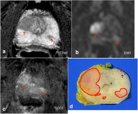
The present study involved 44 patients undergoing 3.0-Tesla multiparametric MRI before laparoscopic radical prostatectomy. Tracing based on pathological and MRI findings was performed. The relationship between the probability of cancer scaled using the 5-point Likert system and the biological characteristics of corresponding tumor foci was evaluated.

RESULTS

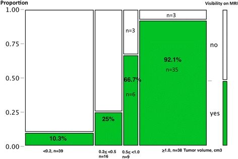
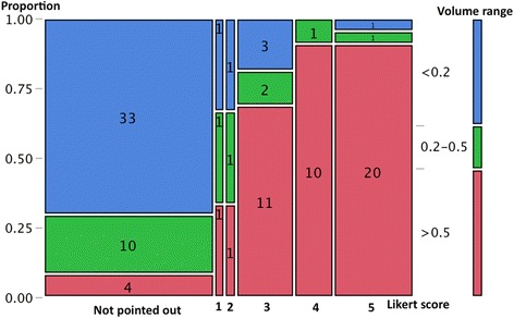
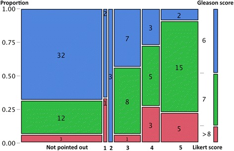
A total of 102 tumor foci were identified histologically from the 44 specimens. Of the 102 tumors, 55 were assigned a score based on MRI findings (score 1: n = 3; score 2: n = 3; score 3: n = 16; score 4: n = 11 score 5: n = 22), while 47 were not pointed out on MRI. The tracing study revealed that the proportion of >0.5 cm(3) tumors increased according to the upgrade of Likert scores (score 1 or 2: 33%; score 3: 68.8%; score 4 or 5: 90.9%, χ(2) test, p < 0.0001). The proportion with a Gleason score >7 also increased from scale 2 to scale 5 (scale 2: 0%; scale 3: 56.3%; scale 4: 72.7%; 5: 90.9%, χ(2) test, p = 0.0001). On using score 3 or higher as the threshold of cancer detection on MRI, the detection rate markedly improved if the tumor volume exceeded 0.5 cm(3) (<0.2 cm(3): 10.3%; 0.2-0.5 cm(3): 25%; 0.5-1.0 cm(3): 66.7%; 1.0 < cm(3): 92.1%).

CONCLUSIONS

Each Likert scale favobably reflected the corresponding tumor's volume and Gleason score. Our observations show that "score 3 or higher" could be a useful threshold to predict clinically significant carcinoma when considering treatment options.

摘要

背景

明确采用5分制李克特量表对前列腺癌可能性进行评分与相应肿瘤病灶生物学特征之间的关系。

方法

本研究纳入44例在腹腔镜前列腺癌根治术前接受3.0T多参数磁共振成像(MRI)检查的患者。基于病理和MRI检查结果进行追踪。评估采用5分制李克特量表对癌症可能性进行评分与相应肿瘤病灶生物学特征之间的关系。

结果

从44份标本中组织学鉴定出共102个肿瘤病灶。在这102个肿瘤中,55个根据MRI检查结果进行了评分(评分1:n = 3;评分2:n = 3;评分3:n = 16;评分4:n = 11;评分5:n = 22),而47个在MRI上未被指出。追踪研究显示,大于0.5cm³的肿瘤比例随李克特评分的升高而增加(评分1或2:33%;评分3:68.8%;评分4或5:90.9%,χ²检验,p < 0.0001)。Gleason评分>7的比例也从量表2到量表5逐渐增加(量表2:0%;量表3:56.3%;量表4:72.7%;量表5:90.9%,χ²检验,p = 0.0001)。以评分3及以上作为MRI上癌症检测的阈值,当肿瘤体积超过0.5cm³时,检测率显著提高(<0.2cm³:10.3%;0.2 - 0.5cm³:25%;0.5 - 1.0cm³:66.7%;1.0 < cm³:92.1%)。

结论

每个李克特量表都能较好地反映相应肿瘤的体积和Gleason评分。我们的观察结果表明,在考虑治疗方案时,“评分3及以上”可能是预测临床显著癌的有用阈值。

https://cdn.ncbi.nlm.nih.gov/pmc/blobs/a82c/4558639/931c4ab12e6f/12894_2015_87_Fig4_HTML.jpg
https://cdn.ncbi.nlm.nih.gov/pmc/blobs/a82c/4558639/7b6f29269ea6/12894_2015_87_Fig1_HTML.jpg
https://cdn.ncbi.nlm.nih.gov/pmc/blobs/a82c/4558639/6ba3c38f0e5f/12894_2015_87_Fig2_HTML.jpg
https://cdn.ncbi.nlm.nih.gov/pmc/blobs/a82c/4558639/b5bdd59c54cc/12894_2015_87_Fig3_HTML.jpg
https://cdn.ncbi.nlm.nih.gov/pmc/blobs/a82c/4558639/931c4ab12e6f/12894_2015_87_Fig4_HTML.jpg

相似文献

1
Five-point Likert scaling on MRI predicts clinically significant prostate carcinoma.磁共振成像(MRI)的五点李克特量表可预测具有临床意义的前列腺癌。
BMC Urol. 2015 Sep 4;15:91. doi: 10.1186/s12894-015-0087-5.
2
[Real-time MRI/US fusion-guided biopsy improves detection rates of prostate cancer in pre-biopsied patients].[实时磁共振成像/超声融合引导活检提高了先前活检患者前列腺癌的检出率]
Aktuelle Urol. 2014 May;45(3):197-203. doi: 10.1055/s-0034-1375682. Epub 2014 Jun 5.
3
Comparison of interreader reproducibility of the prostate imaging reporting and data system and likert scales for evaluation of multiparametric prostate MRI.前列腺影像报告和数据系统与 Likert 量表评估多参数前列腺 MRI 的读者间可重复性比较。
AJR Am J Roentgenol. 2013 Oct;201(4):W612-8. doi: 10.2214/AJR.12.10173.
4
Multiparametric magnetic resonance imaging for the assessment of extracapsular invasion and other staging parameters in patients with prostate cancer candidates for radical prostatectomy.多参数磁共振成像用于评估拟行根治性前列腺切除术的前列腺癌患者的包膜外侵犯及其他分期参数。
Actas Urol Esp. 2014 Jun;38(5):290-7. doi: 10.1016/j.acuro.2013.11.003. Epub 2014 Jan 1.
5
Periprostatic fat thickness on MRI: correlation with Gleason score in prostate cancer.磁共振成像中前列腺周围脂肪厚度与前列腺癌 Gleason 评分的相关性。
AJR Am J Roentgenol. 2015 Jan;204(1):W43-7. doi: 10.2214/AJR.14.12689.
6
How accurate is multiparametric MR imaging in evaluation of prostate cancer volume?多参数磁共振成像评估前列腺癌体积的准确性如何?
Radiology. 2015 Apr;275(1):144-54. doi: 10.1148/radiol.14140524. Epub 2014 Nov 21.
7
Dynamic contrast enhanced MRI of the prostate: comparison of gadobutrol and Gd-DTPA.前列腺动态对比增强磁共振成像:钆布醇与钆喷酸葡胺的比较
Rofo. 2013 Sep;185(9):862-8. doi: 10.1055/s-0033-1335892. Epub 2013 Jul 25.
8
Multiparametric MRI for prostate cancer localization in correlation to whole-mount histopathology.多参数 MRI 与全组织切片病理检查在前列腺癌定位中的相关性。
J Magn Reson Imaging. 2013 Jun;37(6):1392-401. doi: 10.1002/jmri.23938. Epub 2012 Nov 21.
9
Transition zone prostate cancer: detection and localization with 3-T multiparametric MR imaging.移行区前列腺癌:3T 多参数磁共振成像的检测和定位。
Radiology. 2013 Jan;266(1):207-17. doi: 10.1148/radiol.12120281. Epub 2012 Nov 9.
10
[Pharmacokinetic MRI of the prostate: parameters for differentiating low-grade and high-grade prostate cancer].[前列腺的药代动力学磁共振成像:区分低级别和高级别前列腺癌的参数]
Rofo. 2009 Jun;181(6):536-42. doi: 10.1055/s-0028-1109168. Epub 2009 Apr 7.

引用本文的文献

1
Use of Natural Language Processing to Infer Sites of Metastatic Disease From Radiology Reports at Scale.利用自然语言处理技术从大规模放射学报告中推断转移性疾病部位。
JCO Clin Cancer Inform. 2024 May;8:e2300122. doi: 10.1200/CCI.23.00122.
2
The impact of pre-biopsy MRI and additional testing on prostate cancer screening outcomes: A rapid review.活检前MRI及额外检测对前列腺癌筛查结果的影响:一项快速综述。
BJUI Compass. 2024 Jan 31;5(4):426-438. doi: 10.1002/bco2.321. eCollection 2024 Apr.
3
Comparison of Transoral and Transcervical Ultrasonography with MRI for the Diagnostic Work-Up of Oropharynx Tumors: A Protocol for a Multicenter Clinical Trial (SPOTUS).

本文引用的文献

1
Transition zone prostate cancer: revisiting the role of multiparametric MRI at 3 T.移行区前列腺癌:重新探讨 3T 多参数 MRI 的作用。
AJR Am J Roentgenol. 2015 Mar;204(3):W266-72. doi: 10.2214/AJR.14.12955.
2
Prostate Imaging Reporting and Data System and Likert Scoring System: Multiparametric MR Imaging Validation Study to Screen Patients for Initial Biopsy.前列腺影像报告和数据系统和李克特评分系统:多参数磁共振成像验证研究以筛查初始活检患者。
Radiology. 2015 May;275(2):458-68. doi: 10.1148/radiol.14140184. Epub 2015 Jan 19.
3
Evaluation of the PI-RADS scoring system for mpMRI of the prostate: a whole-mount step-section analysis.
经口超声和经颈超声与MRI用于口咽肿瘤诊断检查的比较:一项多中心临床试验方案(SPOTUS)
Diagnostics (Basel). 2024 Mar 8;14(6):577. doi: 10.3390/diagnostics14060577.
4
Differentiating False Positive Lesions from Clinically Significant Cancer and Normal Prostate Tissue Using VERDICT MRI and Other Diffusion Models.使用VERDICT MRI和其他扩散模型区分假阳性病变与具有临床意义的癌症及正常前列腺组织。
Diagnostics (Basel). 2022 Jul 5;12(7):1631. doi: 10.3390/diagnostics12071631.
5
Artificial Intelligence Compared to Radiologists for the Initial Diagnosis of Prostate Cancer on Magnetic Resonance Imaging: A Systematic Review and Recommendations for Future Studies.人工智能与放射科医生在磁共振成像上对前列腺癌进行初步诊断的比较:一项系统评价及对未来研究的建议
Cancers (Basel). 2021 Jul 1;13(13):3318. doi: 10.3390/cancers13133318.
6
A Radiogenomic Approach for Decoding Molecular Mechanisms Underlying Tumor Progression in Prostate Cancer.一种用于解码前列腺癌肿瘤进展潜在分子机制的放射基因组学方法。
Cancers (Basel). 2019 Sep 2;11(9):1293. doi: 10.3390/cancers11091293.
7
The Oncogene Metadherin Interacts with the Known Splicing Proteins YTHDC1, Sam68 and T-STAR and Plays a Novel Role in Alternative mRNA Splicing.致癌基因间黏附分子与已知的剪接蛋白YTHDC1、Sam68和T-STAR相互作用,并在可变mRNA剪接中发挥新作用。
Cancers (Basel). 2019 Aug 23;11(9):1233. doi: 10.3390/cancers11091233.
8
Optimization and repeatability of multipool chemical exchange saturation transfer MRI of the prostate at 3.0 T.3.0T 多池化学交换饱和传递 MRI 前列腺的优化和可重复性。
J Magn Reson Imaging. 2019 Oct;50(4):1238-1250. doi: 10.1002/jmri.26690. Epub 2019 Feb 15.
9
Prostate imaging features that indicate benign or malignant pathology on biopsy.前列腺成像特征可在活检时提示良性或恶性病变。
Transl Androl Urol. 2018 Sep;7(Suppl 4):S420-S435. doi: 10.21037/tau.2018.07.06.
10
Imaging for the selection and monitoring of men on active surveillance for prostate cancer.用于前列腺癌主动监测男性患者的选择和监测的影像学检查。
Transl Androl Urol. 2018 Apr;7(2):228-235. doi: 10.21037/tau.2017.08.13.
前列腺磁共振成像(mpMRI)的PI-RADS评分系统评估:全层阶梯切片分析
World J Urol. 2015 Jul;33(7):1023-30. doi: 10.1007/s00345-014-1370-x. Epub 2014 Aug 1.
4
Comparison of endorectal coil and nonendorectal coil T2W and diffusion-weighted MRI at 3 Tesla for localizing prostate cancer: correlation with whole-mount histopathology.3特斯拉下直肠内线圈与非直肠内线圈T2加权成像及扩散加权磁共振成像在前列腺癌定位中的比较:与全层组织病理学的相关性
J Magn Reson Imaging. 2014 Jun;39(6):1443-8. doi: 10.1002/jmri.24317. Epub 2013 Nov 15.
5
Prostate cancer localization using multiparametric MR imaging: comparison of Prostate Imaging Reporting and Data System (PI-RADS) and Likert scales.多参数磁共振成像在前列腺癌定位中的应用:前列腺影像报告和数据系统(PI-RADS)与李克特量表的比较。
Radiology. 2013 Nov;269(2):482-92. doi: 10.1148/radiol.13122233. Epub 2013 Jun 20.
6
Scoring systems used for the interpretation and reporting of multiparametric MRI for prostate cancer detection, localization, and characterization: could standardization lead to improved utilization of imaging within the diagnostic pathway?用于解释和报告前列腺癌检测、定位和特征的多参数 MRI 的评分系统:标准化是否能提高诊断途径中影像学的应用?
J Magn Reson Imaging. 2013 Jan;37(1):48-58. doi: 10.1002/jmri.23689. Epub 2012 May 7.
7
Comparison of pelvic phased-array versus endorectal coil magnetic resonance imaging at 3 Tesla for local staging of prostate cancer.3T 下盆腔相控阵与直肠内线圈磁共振成像在前列腺癌局部分期中的比较。
Yonsei Med J. 2012 May;53(3):550-6. doi: 10.3349/ymj.2012.53.3.550.
8
ESUR prostate MR guidelines 2012.ESUR 前列腺磁共振成像指南 2012.
Eur Radiol. 2012 Apr;22(4):746-57. doi: 10.1007/s00330-011-2377-y. Epub 2012 Feb 10.
9
Multiparametric 3T prostate magnetic resonance imaging to detect cancer: histopathological correlation using prostatectomy specimens processed in customized magnetic resonance imaging based molds.多参数 3T 前列腺磁共振成像检测癌症:使用基于定制磁共振成像模具处理的前列腺切除术标本进行组织病理学相关性研究。
J Urol. 2011 Nov;186(5):1818-24. doi: 10.1016/j.juro.2011.07.013. Epub 2011 Sep 25.
10
Diffusion-weighted endorectal MR imaging at 3 T for prostate cancer: tumor detection and assessment of aggressiveness.3T 直肠内磁共振弥散加权成像在前列腺癌中的应用:肿瘤检测及侵袭性评估。
Radiology. 2011 Jun;259(3):775-84. doi: 10.1148/radiol.11102066. Epub 2011 Mar 24.