Shi Jianhua, Gu Jin-hua, Dai Chun-ling, Gu Jianlan, Jin Xiaoxia, Sun Jianming, Iqbal Khalid, Liu Fei, Gong Cheng-Xin
Jiangsu Key Laboratory of Neuroregeneration, Co-Innovation Center of Neuroregeneration, Nantong University, Nantong, Jiangsu 226001, China.
Department of Biochemistry, Nantong University Medical School, Nantong, Jiangsu 226001, China.
Sci Rep. 2015 Sep 28;5:14500. doi: 10.1038/srep14500.
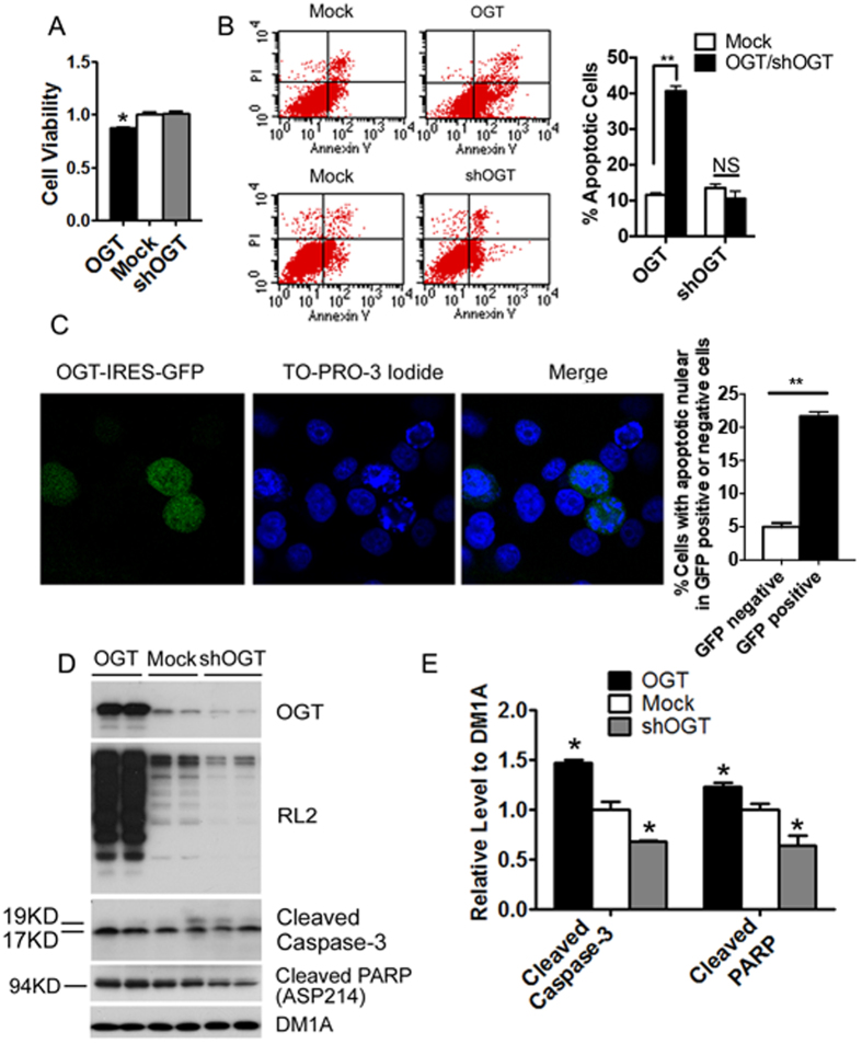
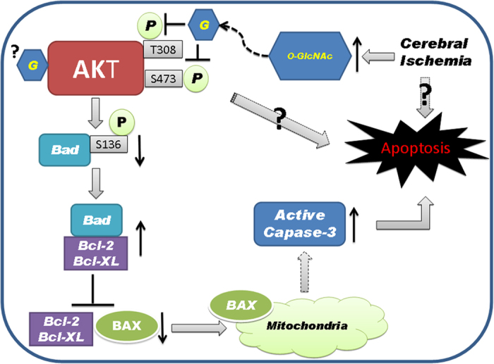
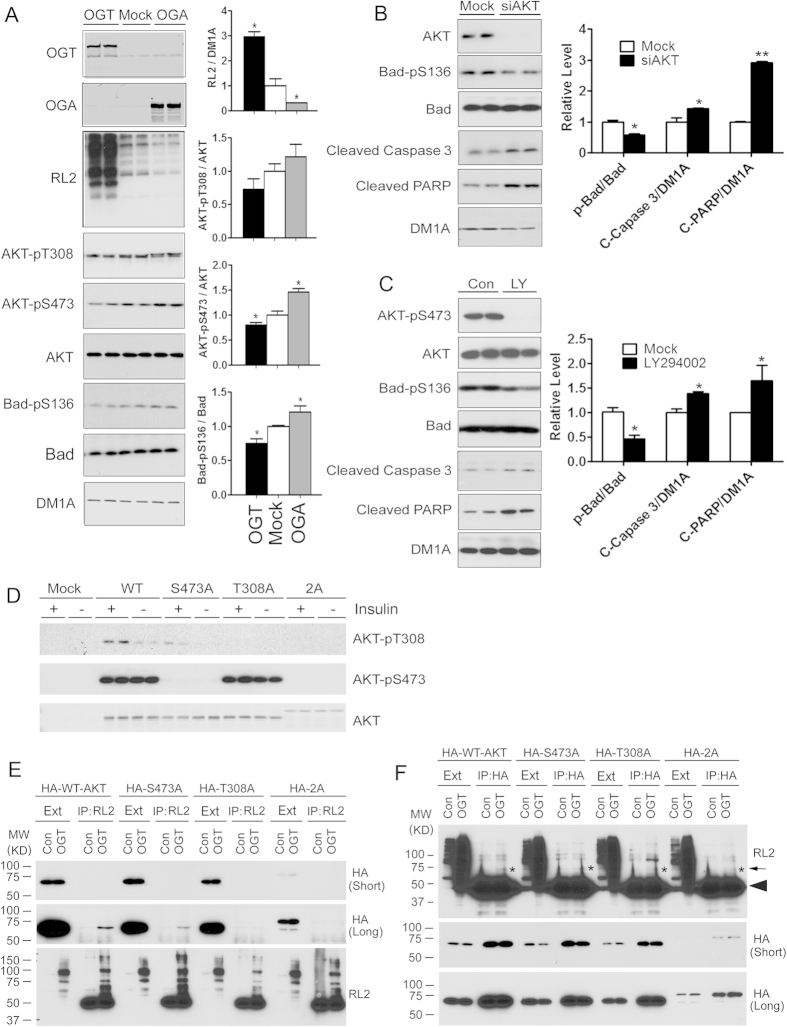
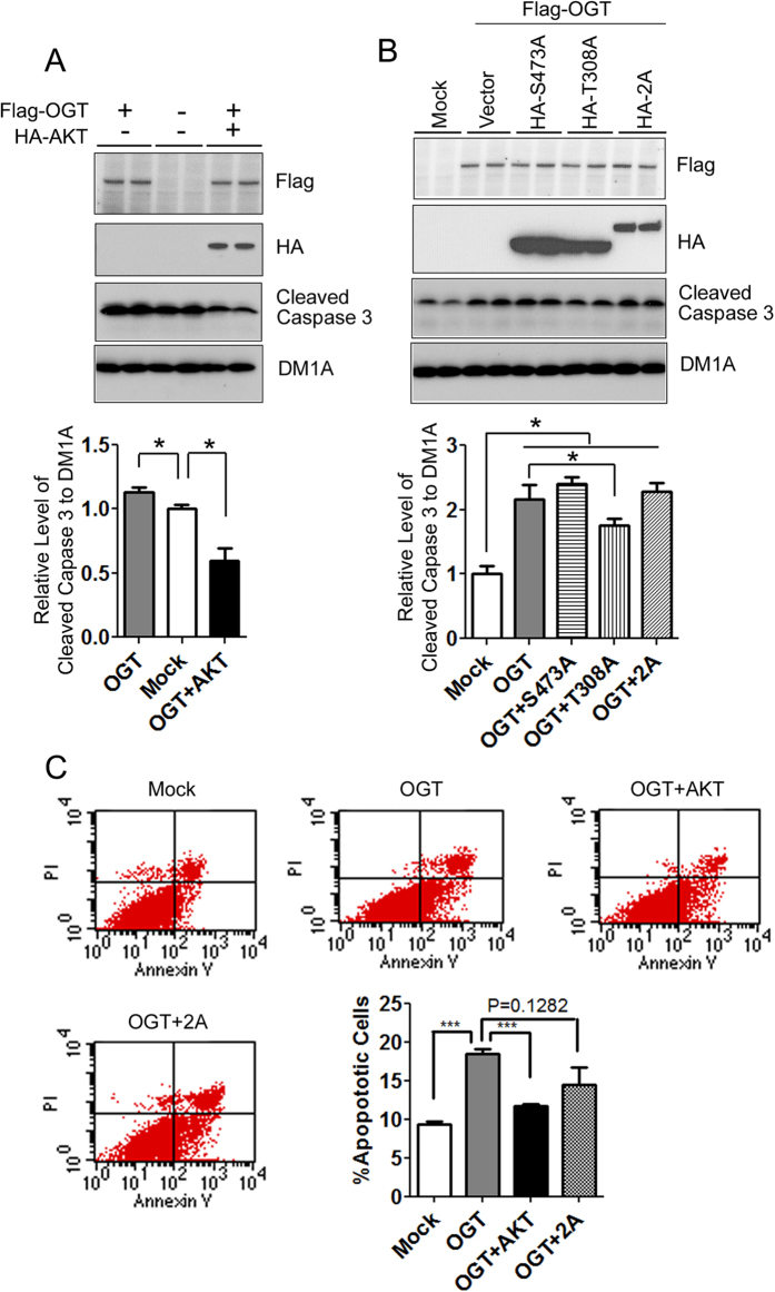
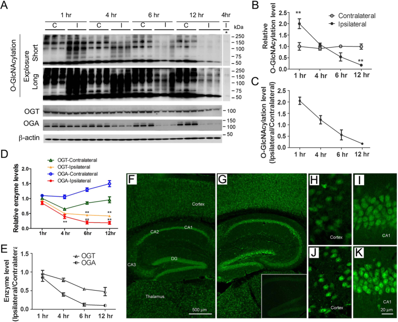
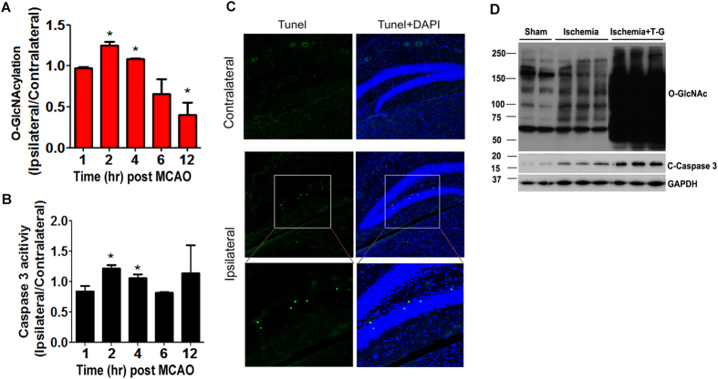
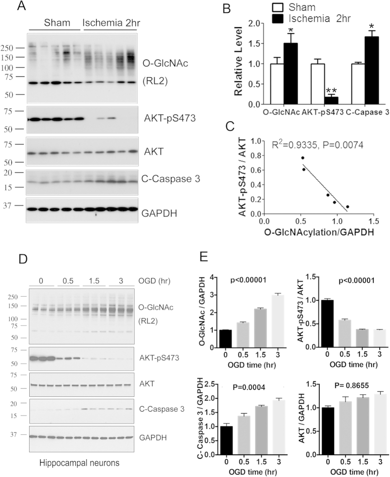
Apoptosis plays an important role in neural development and neurological disorders. In this study, we found that O-GlcNAcylation, a unique protein posttranslational modification with O-linked β-N-acetylglucosamine (GlcNAc), promoted apoptosis through attenuating phosphorylation/activation of AKT and Bad. By using co-immunoprecipitation and mutagenesis techniques, we identified O-GlcNAc modification at both Thr308 and Ser473 of AKT. O-GlcNAcylation-induced apoptosis was attenuated by over-expression of AKT. We also found a dynamic elevation of protein O-GlcNAcylation during the first four hours of cerebral ischemia, followed by continuous decline after middle cerebral artery occlusion (MCAO) in the mouse brain. The elevation of O-GlcNAcylation coincided with activation of cell apoptosis. Finally, we found a negative correlation between AKT phosphorylation and O-GlcNAcylation in ischemic brain tissue. These results indicate that cerebral ischemia induces a rapid increase of O-GlcNAcylation that promotes apoptosis through down-regulation of AKT activity. These findings provide a novel mechanism through which O-GlcNAcylation regulates ischemia-induced neuronal apoptosis through AKT signaling.
细胞凋亡在神经发育和神经疾病中发挥着重要作用。在本研究中,我们发现O-连接的β-N-乙酰葡糖胺(GlcNAc)这种独特的蛋白质翻译后修饰——O-GlcNAc糖基化,通过减弱AKT和Bad的磷酸化/激活来促进细胞凋亡。通过使用免疫共沉淀和诱变技术,我们鉴定出AKT的苏氨酸308和丝氨酸473位点均存在O-GlcNAc修饰。过表达AKT可减弱O-GlcNAc糖基化诱导的细胞凋亡。我们还发现,在小鼠大脑大脑中动脉闭塞(MCAO)后,脑缺血的最初四小时内蛋白质O-GlcNAc糖基化呈动态升高,随后持续下降。O-GlcNAc糖基化的升高与细胞凋亡的激活同时发生。最后,我们发现缺血脑组织中AKT磷酸化与O-GlcNAc糖基化呈负相关。这些结果表明,脑缺血诱导O-GlcNAc糖基化迅速增加,通过下调AKT活性促进细胞凋亡。这些发现提供了一种新的机制,通过该机制O-GlcNAc糖基化通过AKT信号通路调节缺血诱导的神经元凋亡。