Nguyen Ha Son, Doan Ninh, Eckardt Gerald, Gelsomino Michael, Shabani Saman, Brown W Douglas, Mueller Wade, Pollock Glen
Department of Neurosurgery, Medical College of Wisconsin, Milwaukee, WI, USA.
Department of Radiology, Medical College of Wisconsin, Milwaukee, WI, USA.
Surg Neurol Int. 2015 Sep 7;6:146. doi: 10.4103/2152-7806.164696. eCollection 2015.
Few reports exist regarding thrombosed aneurysms where the initial work up was concerning for a neoplasm. To date, no published reports exist regarding a nongiant thrombosed middle cerebral artery aneurysm, where the primary workup and treatment plan was directed toward a preliminary diagnosis of intra-axial neoplasm.
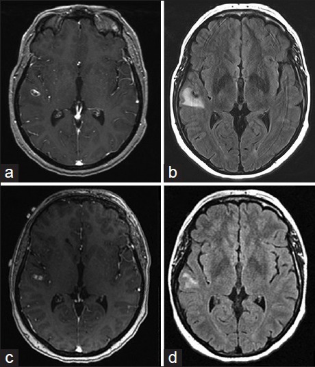
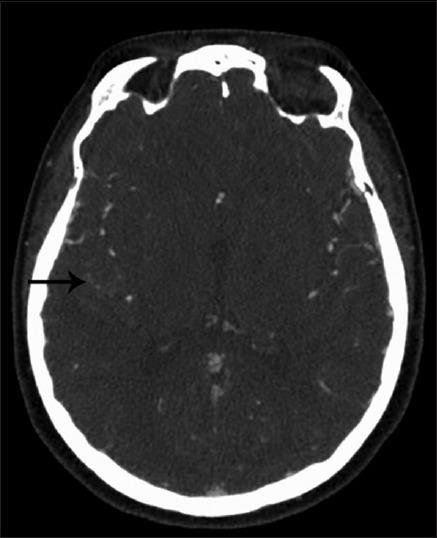
We report a 43-year-old female who presented with a generalized tonic-clonic seizure attributed to a lesion along the right superior temporal gyrus. The lesion enhanced on initial magnetic resonance imaging (MRI) of the brain, as well as on follow-up MRI. Subsequent vascular studies and metastatic work up were negative. A craniotomy with image guidance was performed and an intraoperative diagnosis was made of a thrombosed aneurysm along a branch of the middle cerebral artery. The aneurysm was trapped and resected as there was no significant flow from the branch as seen on the prior cerebral angiogram. The patient had an uneventful postoperative course.
Completely thrombosed, nongiant aneurysms can mimic an intra-axial neoplasm. Typical imaging features for thrombosed aneurysms may be missed, especially if the aneurysms are small, where imaging characteristics of the intraluminal contents is more difficult to appreciate. Although imaging may be consistent with a neoplastic lesion, there should be suspicion for a potential underlying aneurysm.
关于最初检查怀疑为肿瘤的血栓性动脉瘤的报道很少。迄今为止,尚无关于非巨大血栓性大脑中动脉动脉瘤的已发表报道,其主要检查和治疗计划是针对轴内肿瘤的初步诊断。
我们报告一名43岁女性,因右侧颞上回病变出现全身强直阵挛性发作。该病变在初次脑部磁共振成像(MRI)以及随访MRI上均有强化。随后的血管检查和转移灶检查均为阴性。在图像引导下进行了开颅手术,术中诊断为大脑中动脉分支处的血栓性动脉瘤。由于在之前的脑血管造影中未见该分支有明显血流,故将动脉瘤夹闭并切除。患者术后恢复顺利。
完全血栓形成的非巨大动脉瘤可酷似轴内肿瘤。血栓性动脉瘤的典型影像学特征可能会被遗漏,尤其是当动脉瘤较小,管腔内内容物的影像学特征更难辨认时。尽管影像学表现可能与肿瘤性病变一致,但仍应怀疑潜在的动脉瘤。