Mollink J, van Baarsen K M, Dederen P J W C, Foxley S, Miller K L, Jbabdi S, Slump C H, Grotenhuis J A, Kleinnijenhuis M, van Cappellen van Walsum A M
Nuffield Department of Clinical Neurosciences, FMRIB Centre, University of Oxford, Oxford, UK.
Department of Anatomy, Donders Institute for Brain Cognition and Behaviour, Radboud University Medical Centre, Nijmegen, The Netherlands.
Brain Struct Funct. 2016 Sep;221(7):3487-501. doi: 10.1007/s00429-015-1115-7. Epub 2015 Oct 5.
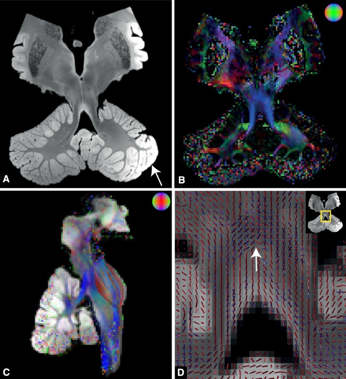
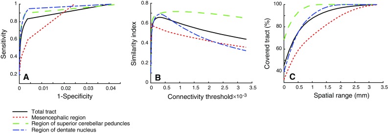
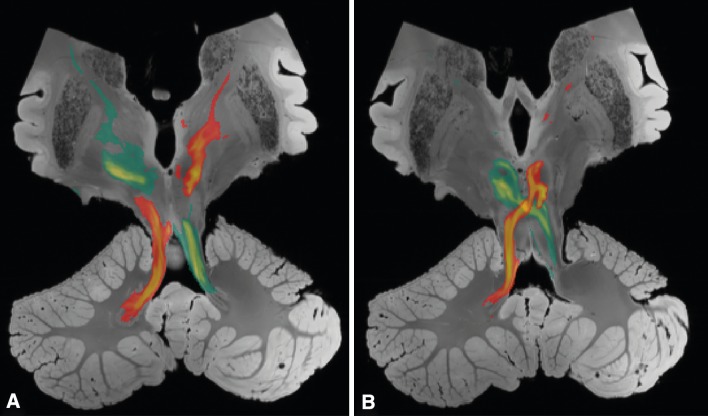
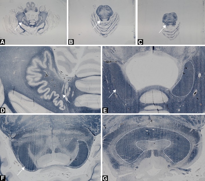
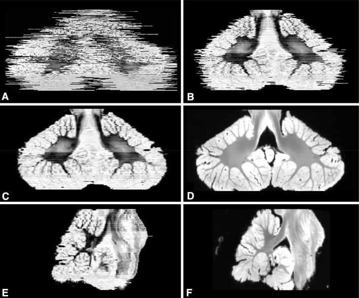
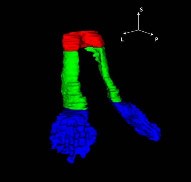
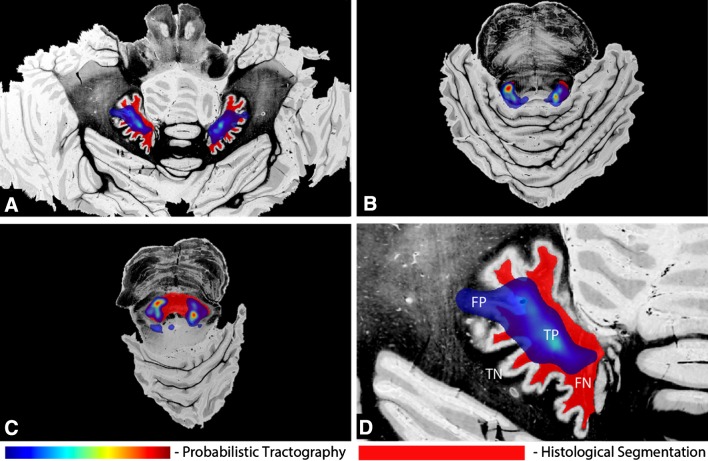
Diffusion-weighted imaging (DWI) tractography is a technique with great potential to characterize the in vivo anatomical position and integrity of white matter tracts. Tractography, however, remains an estimation of white matter tracts, and false-positive and false-negative rates are not available. The goal of the present study was to compare postmortem tractography of the dentatorubrothalamic tract (DRTT) by its 3D histological reconstruction, to estimate the reliability of the tractography algorithm in this specific tract. Recent studies have shown that the cerebellum is involved in cognitive, language and emotional functions besides its role in motor control. However, the exact working mechanism of the cerebellum is still to be elucidated. As the DRTT is the main output tract it is of special interest for the neuroscience and clinical community. A postmortem human brain specimen was scanned on a 7T MRI scanner using a diffusion-weighted steady-state free precession sequence. Tractography was performed with PROBTRACKX. The specimen was subsequently serially sectioned and stained for myelin using a modified Heidenhain-Woelke staining. Image registration permitted the 3D reconstruction of the histological sections and comparison with MRI. The spatial concordance between the two modalities was evaluated using ROC analysis and a similarity index (SI). ROC curves showed a high sensitivity and specificity in general. Highest measures were observed in the superior cerebellar peduncle with an SI of 0.72. Less overlap was found in the decussation of the DRTT at the level of the mesencephalon. The study demonstrates high spatial accuracy of postmortem probabilistic tractography of the DRTT when compared to a 3D histological reconstruction. This gives hopeful prospect for studying structure-function correlations in patients with cerebellar disorders using tractography of the DRTT.
扩散加权成像(DWI)纤维束成像技术在表征白质纤维束的体内解剖位置和完整性方面具有巨大潜力。然而,纤维束成像仍然是对白质纤维束的一种估计,且假阳性和假阴性率尚不可知。本研究的目的是通过齿状红核丘脑束(DRTT)的三维组织学重建来比较其死后纤维束成像,以评估该特定纤维束成像算法的可靠性。最近的研究表明,小脑除了在运动控制中发挥作用外,还参与认知、语言和情感功能。然而,小脑的确切工作机制仍有待阐明。由于DRTT是主要的输出纤维束,因此它对神经科学和临床领域具有特殊意义。使用扩散加权稳态自由进动序列在7T MRI扫描仪上对一个死后人类脑标本进行扫描。使用PROBTRACKX进行纤维束成像。随后对标本进行连续切片,并使用改良的海登海因 - 韦尔克染色法对髓磷脂进行染色。图像配准允许对组织学切片进行三维重建并与MRI进行比较。使用ROC分析和相似性指数(SI)评估两种模式之间的空间一致性。ROC曲线总体上显示出高敏感性和特异性。在小脑上脚观察到最高测量值,SI为0.72。在中脑水平的DRTT交叉处发现的重叠较少。该研究表明,与三维组织学重建相比,DRTT死后概率性纤维束成像具有较高的空间准确性。这为使用DRTT纤维束成像研究小脑疾病患者的结构 - 功能相关性提供了充满希望的前景。