Castellaro Andrés M, Tonda Alfredo, Cejas Hugo H, Ferreyra Héctor, Caputto Beatriz L, Pucci Oscar A, Gil German A
Departamento de Química Biológica, Facultad de Ciencias Químicas, Universidad Nacional de Córdoba- CIQUIBIC, CONICET, Córdoba, Argentina.
Primera Cátedra de Ginecología, Hospital Nacional de Clínicas, Universidad Nacional de Córdoba, Córdoba, Argentina.
BMC Cancer. 2015 Oct 22;15:761. doi: 10.1186/s12885-015-1747-2.
Microcalcifications can be the early and only presenting sign of breast cancer. One shared characteristic of breast cancer is the appearance of mammographic mammary microcalcifications that can routinely be used to detect breast cancer in its initial stages, which is of key importance due to the possibility that early detection allows the application of more conservative therapies for a better patient outcome. The mechanism by which mammary microcalcifications are formed is still largely unknown but breast cancers presenting microcalcifications are more often associated with a poorer prognosis.
We combined Capillary Electrochromatography, histology, and gene expression (qRT-PCR) to analyze patient-matched normal breast tissue vs. breast tumor. Potential carcinogenicity of oxalate was tested by its inoculation into mice. All data were subjected to statistical analysis.
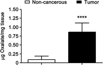
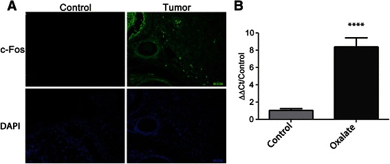
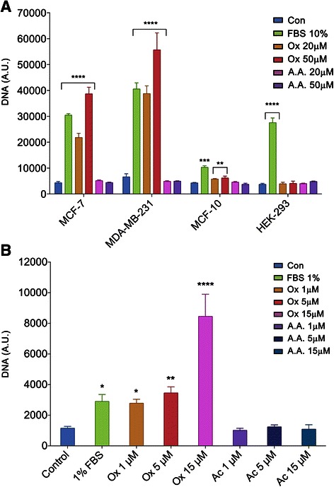
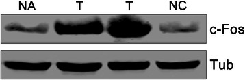
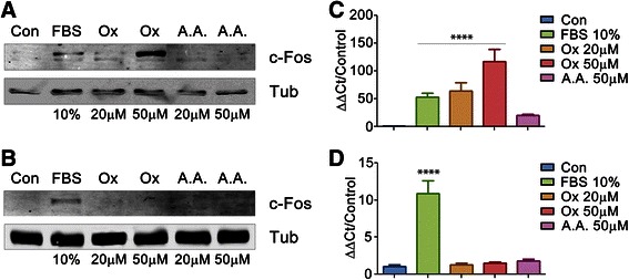
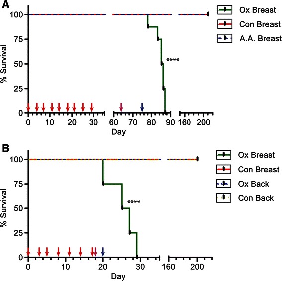
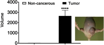
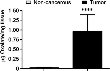
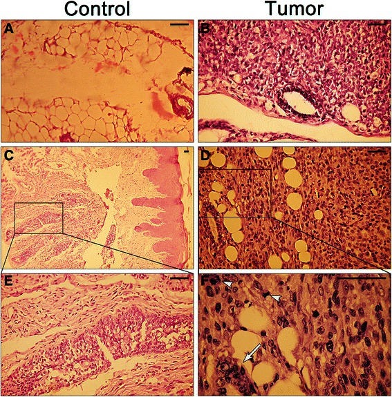
To study the biological significance of oxalates within the breast tumor microenvironment, we measured oxalate concentration in both human breast tumor tissues and adjoining non-pathological breast tissues. We found that all tested breast tumor tissues contain a higher concentration of oxalates than their counterpart non-pathological breast tissue. Moreover, it was established that oxalate induces proliferation of breast cells and stimulates the expression of a pro-tumorigenic gene c-fos. Furthermore, oxalate generates highly malignant and undifferentiated tumors when it was injected into the mammary fatpad in female mice, but not when injected into their back, indicating that oxalate does not induce cancer formation in all types of tissues. Moreover, neither human kidney-epithelial cells nor mouse fibroblast cells proliferate when are treated with oxalate.
We found that the chronic exposure of breast epithelial cells to oxalate promotes the transformation of breast cells from normal to tumor cells, inducing the expression of a proto-oncogen as c-fos and proliferation in breast cancer cells. Furthermore, oxalate has a carcinogenic effect when injected into the mammary fatpad in mice, generating highly malignant and undifferentiated tumors with the characteristics of fibrosarcomas of the breast. As oxalates seem to promote these differences, it is expected that a significant reduction in the incidence of breast cancer tumors could be reached if it were possible to control oxalate production or its carcinogenic activity.
微钙化可能是乳腺癌早期唯一的表现迹象。乳腺癌的一个共同特征是乳腺钼靶微钙化的出现,这可常规用于在乳腺癌的初始阶段进行检测,鉴于早期检测有可能使更保守的治疗得以应用从而获得更好的患者预后,这一点至关重要。乳腺微钙化形成的机制在很大程度上仍不清楚,但出现微钙化的乳腺癌往往预后较差。
我们结合毛细管电色谱、组织学和基因表达(定量逆转录聚合酶链反应)来分析患者匹配的正常乳腺组织与乳腺肿瘤。通过将草酸盐接种到小鼠体内来测试其潜在致癌性。所有数据均进行统计分析。
为研究草酸盐在乳腺肿瘤微环境中的生物学意义,我们测量了人乳腺肿瘤组织和相邻非病理乳腺组织中的草酸盐浓度。我们发现,所有测试的乳腺肿瘤组织中草酸盐浓度均高于其对应的非病理乳腺组织。此外,已证实草酸盐可诱导乳腺细胞增殖并刺激促肿瘤基因c-fos的表达。此外,当将草酸盐注射到雌性小鼠的乳腺脂肪垫中时会产生高度恶性且未分化的肿瘤,但注射到其背部时则不会,这表明草酸盐并非在所有类型的组织中都能诱导癌症形成。而且,用草酸盐处理时,人肾上皮细胞和小鼠成纤维细胞均不会增殖。
我们发现,乳腺上皮细胞长期暴露于草酸盐会促使乳腺细胞从正常细胞转变为肿瘤细胞,诱导原癌基因如c-fos的表达并使乳腺癌细胞增殖。此外,将草酸盐注射到小鼠乳腺脂肪垫中时具有致癌作用,会产生具有乳腺纤维肉瘤特征的高度恶性且未分化的肿瘤。由于草酸盐似乎会促成这些差异,因此如果能够控制草酸盐的产生或其致癌活性,预计乳腺癌肿瘤的发病率可能会显著降低。