Tang Wenjing, Song Huanlei, Cai Wei, Shen Xiuhua
Department of Clinical Nutrition, Xin Hua Hospital, School of Medicine, Shanghai Jiao Tong University, Shanghai 200092, China.
Department of Nutrition, School of Medicine, Shanghai Jiao Tong University, Shanghai 200025, China.
Nutrients. 2015 Oct 27;7(10):8871-86. doi: 10.3390/nu7105437.
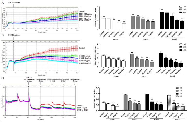
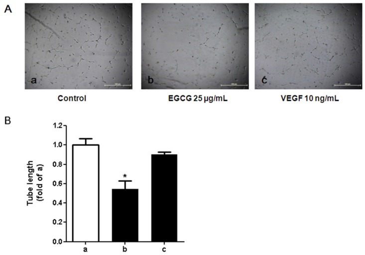
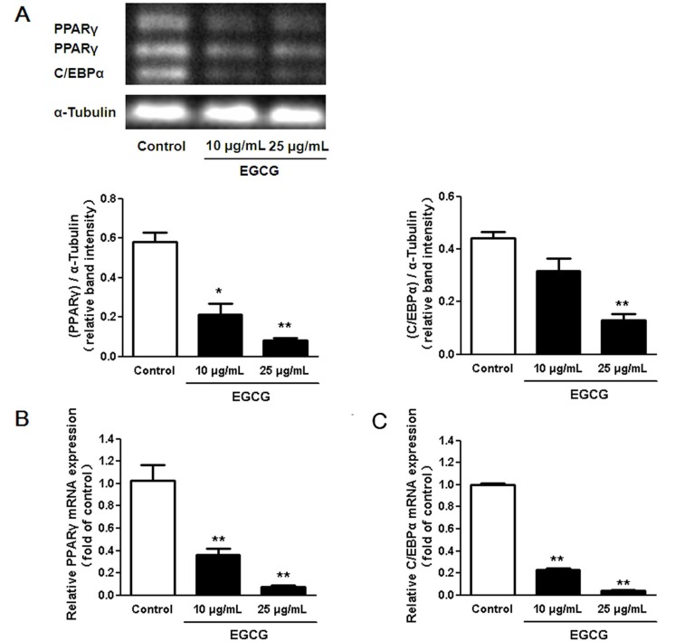
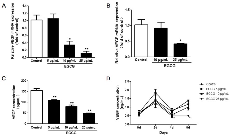
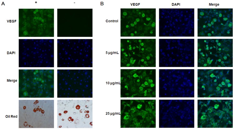
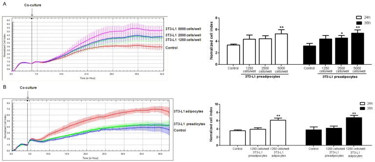
Little is known about the effect of (-)-epigallocatechin-3-gallate (EGCG) on angiogenesis in adipocytes. We aimed to test the effect of EGCG on the expression of vascular endothelial growth factor (VEGF) in adipocytes. The levels of VEGF secretion, the expression of VEGF message ribonucleic acid (mRNA) and VEGF protein in 3T3-L1 cells were measured by enzyme linked immunosorbent assay (ELISA), real time polymerase chain reaction (PCR), and immunofluorescence staining, respectively. The xCELLigence real time cell analysis system was used to study the growth and differentiation of 3T3-L1 preadipocytes. A coculture system was used to test the effects of 3T3-L1 cells on proliferation of human umbilical vein endothelial cells (HUVECs). The conditioned media derived from 3T3-L1 cells treated with or without EGCG was used to culture the HUVECs for a tube formation assay. Peroxisome proliferator-activated receptor γ (PPARγ) and CCAAT/enhancer binding protein α (C/EBPα), two transcription factors related to both adipogenesis and angiogenesis, were examined to explore the potential mechanism. We found that all the three measurements of VEGF expression in adipocytes (mRNA, protein and secretion in media) were reduced after EGCG treatment. The growth of HUVECs co-cultured with 3T3-L1 cells was significantly increased and the conditioned media from EGCG treated 3T3-L1 adipocytes inhibited tube formation in HUVECs. Both PPARγ and C/EBPα expression in adipocytes were decreased with EGCG treatment. In conclusion, findings from this study suggest that EGCG may inhibit angiogenesis by regulating VEGF expression and secretion in adipocytes.
关于(-)-表没食子儿茶素-3-没食子酸酯(EGCG)对脂肪细胞血管生成的影响,目前所知甚少。我们旨在测试EGCG对脂肪细胞中血管内皮生长因子(VEGF)表达的影响。分别通过酶联免疫吸附测定(ELISA)、实时聚合酶链反应(PCR)和免疫荧光染色,检测3T3-L1细胞中VEGF的分泌水平、VEGF信使核糖核酸(mRNA)和VEGF蛋白的表达。使用xCELLigence实时细胞分析系统研究3T3-L1前脂肪细胞的生长和分化。采用共培养系统测试3T3-L1细胞对人脐静脉内皮细胞(HUVECs)增殖的影响。用含或不含EGCG处理的3T3-L1细胞的条件培养基培养HUVECs,进行管形成试验。检测过氧化物酶体增殖物激活受体γ(PPARγ)和CCAAT/增强子结合蛋白α(C/EBPα)这两种与脂肪生成和血管生成相关的转录因子,以探讨潜在机制。我们发现,EGCG处理后,脂肪细胞中VEGF表达的所有三项测量指标(mRNA、蛋白和培养基中的分泌)均降低。与3T3-L1细胞共培养的HUVECs的生长显著增加,而经EGCG处理的3T3-L1脂肪细胞的条件培养基抑制了HUVECs的管形成。EGCG处理使脂肪细胞中PPARγ和C/EBPα的表达均降低。总之,本研究结果表明,EGCG可能通过调节脂肪细胞中VEGF的表达和分泌来抑制血管生成。