Shin Hyeon-Jun, Kwon Hyuk-Kwon, Lee Jae-Hyeok, Gui Xiangai, Achek Asma, Kim Jae-Ho, Choi Sangdun
Department of Molecular Science and Technology, Ajou University, Suwon 443-749, Korea.
Sci Rep. 2015 Nov 2;5:15798. doi: 10.1038/srep15798.
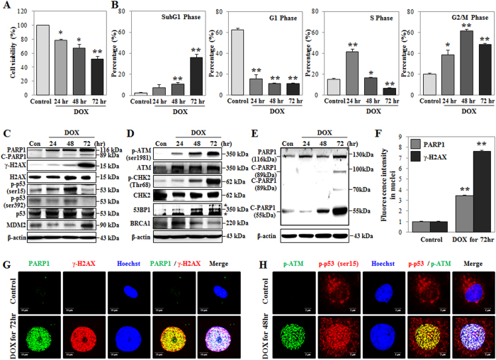
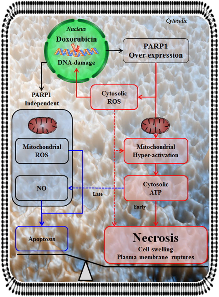
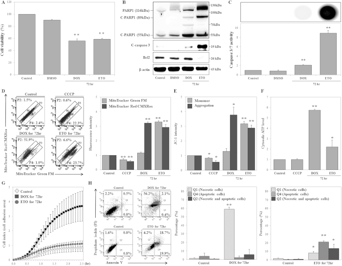
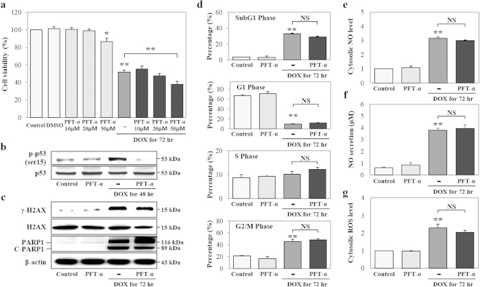
Necrosis, unregulated cell death, is characterized by plasma membrane rupture as well as nuclear and cellular swelling. However, it has recently been reported that necrosis is a regulated form of cell death mediated by poly-(ADP-ribose) polymerase 1 (PARP1). PARP1 is thought to mediate necrosis by inducing DNA damage, although this remains unconfirmed. In this study, we examined the mechanisms of PARP1-mediated necrosis following doxorubicin (DOX)-induced DNA damage in human kidney proximal tubular (HK-2) cells. DOX initiated DNA damage response (DDR) and upregulated PARP1 and p53 expression, resulting in morphological changes similar to those observed during necrosis. Additionally, DOX induced mitochondrial hyper-activation, as evidenced by increased mitochondrial respiration and cytosolic ATP (cATP) production. However, DOX affected mitochondrial mass. DOX-induced DNA damage, cytosolic reactive oxygen species (cROS) generation, and mitochondrial hyper-activation decreased in cells with inhibited PARP1 expression, while generation of nitric oxide (NO) and mitochondrial ROS (mROS) remained unaffected. Moreover, DOX-induced DNA damage, cell cycle changes, and oxidative stress were not affected by p53 inhibition. These findings suggest that DNA damage induced necrosis through a PARP1-dependent and p53-independent pathway.
坏死,即不受调控的细胞死亡,其特征为质膜破裂以及细胞核和细胞肿胀。然而,最近有报道称坏死是一种由聚(ADP - 核糖)聚合酶1(PARP1)介导的受调控的细胞死亡形式。尽管这一点尚未得到证实,但PARP1被认为通过诱导DNA损伤来介导坏死。在本研究中,我们检测了阿霉素(DOX)诱导人肾近端小管(HK - 2)细胞DNA损伤后PARP1介导坏死的机制。DOX引发了DNA损伤反应(DDR)并上调了PARP1和p53的表达,导致出现与坏死过程中观察到的类似形态变化。此外,DOX诱导线粒体过度激活,线粒体呼吸增加和胞质ATP(cATP)生成增加证明了这一点。然而,DOX影响线粒体质量。在PARP1表达受到抑制的细胞中,DOX诱导的DNA损伤、胞质活性氧(cROS)生成和线粒体过度激活减少,而一氧化氮(NO)和线粒体ROS(mROS)的生成不受影响。此外,DOX诱导的DNA损伤、细胞周期变化和氧化应激不受p53抑制的影响。这些发现表明,DNA损伤通过PARP1依赖性和p53非依赖性途径诱导坏死。