Farjam Reza, Pramanik Priyanka, Aryal Madhava P, Srinivasan Ashok, Chapman Christopher H, Tsien Christina I, Lawrence Theodore S, Cao Yue
Department of Medical Physics, Memorial Sloan-Kettering Cancer Center, New York, New York.
Department of Radiation Oncology, University of Michigan, Ann Arbor, Michigan.
Int J Radiat Oncol Biol Phys. 2015 Nov 15;93(4):908-15. doi: 10.1016/j.ijrobp.2015.08.014. Epub 2015 Aug 8.
We aimed to develop a hippocampal vascular injury surrogate marker for early prediction of late neurocognitive dysfunction in patients receiving brain radiation therapy (RT).
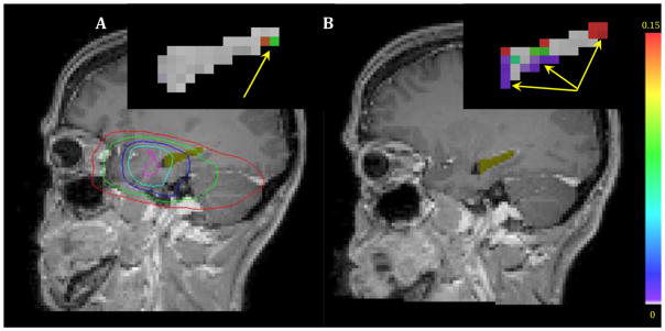
Twenty-seven patients (17 males and 10 females, 31-80 years of age) were enrolled in an institutional review board-approved prospective longitudinal study. Patients received diagnoses of low-grade glioma or benign tumor and were treated by (3D) conformal or intensity-modulated RT with a median dose of 54 Gy (50.4-59.4 Gy in 1.8-Gy fractions). Six dynamic-contrast enhanced MRI scans were performed from pre-RT to 18-month post-RT, and quantified for vascular parameters related to blood-brain barrier permeability, K(trans), and the fraction of blood plasma volume, Vp. The temporal changes in the means of hippocampal transfer constant K(trans) and Vp after starting RT were modeled by integrating the dose effects with age, sex, hippocampal laterality, and presence of tumor or edema near a hippocampus. Finally, the early vascular dose response in hippocampi was correlated with neurocognitive dysfunction at 6 and 18 months post-RT.
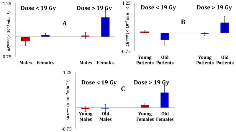
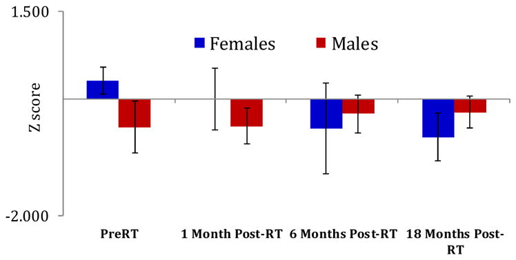
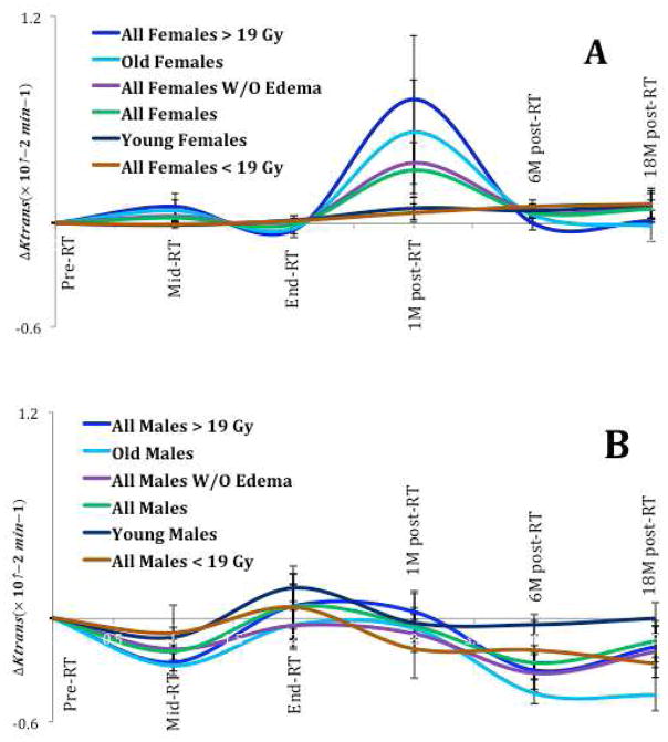
The mean K(trans) Increased significantly from pre-RT to 1-month post-RT (P<.0004), which significantly depended on sex (P<.0007) and age (P<.00004), with the dose response more pronounced in older females. Also, the vascular dose response in the left hippocampus of females correlated significantly with changes in memory function at 6 (r=-0.95, P<.0006) and 18-months (r=-0.88, P<.02) post-RT.
The early hippocampal vascular dose response could be a predictor of late neurocognitive dysfunction. A personalized hippocampus sparing strategy may be considered in the future.
我们旨在开发一种海马体血管损伤替代标志物,用于早期预测接受脑部放射治疗(RT)患者的晚期神经认知功能障碍。
27例患者(17例男性和10例女性,年龄31 - 80岁)纳入一项经机构审查委员会批准的前瞻性纵向研究。患者被诊断为低级胶质瘤或良性肿瘤,并接受(3D)适形或调强放疗,中位剂量为54 Gy(1.8 Gy分割,剂量范围50.4 - 59.4 Gy)。从放疗前至放疗后18个月进行6次动态对比增强MRI扫描,并对与血脑屏障通透性相关的血管参数、转运常数K(trans)和血浆容积分数Vp进行量化。通过整合剂量效应与年龄、性别、海马体侧别以及海马体附近肿瘤或水肿的存在情况,对放疗开始后海马体转运常数K(trans)和Vp均值的时间变化进行建模。最后,将海马体的早期血管剂量反应与放疗后6个月和18个月时的神经认知功能障碍进行相关性分析。
从放疗前至放疗后1个月,平均K(trans)显著增加(P <.0004),这显著取决于性别(P <.0007)和年龄(P <.00004),老年女性的剂量反应更明显。此外,女性左侧海马体的血管剂量反应与放疗后6个月(r = -0.95,P <.0006)和18个月(r = -0.88,P <.02)时记忆功能的变化显著相关。
早期海马体血管剂量反应可能是晚期神经认知功能障碍的预测指标。未来可考虑采用个性化的海马体保护策略。