Eke Iris, Hehlgans Stephanie, Sandfort Veit, Cordes Nils
OncoRay-National Center for Radiation Research in Oncology, Medical Faculty Carl Gustav Carus, Technische Universität Dresden, D-01307 Dresden, Germany.
Department of Radiology and Imaging Sciences, Clinical Center, National Institutes of Health, Bethesda, MD 20892, USA.
Int J Oncol. 2016 Jan;48(1):313-21. doi: 10.3892/ijo.2015.3230. Epub 2015 Nov 4.
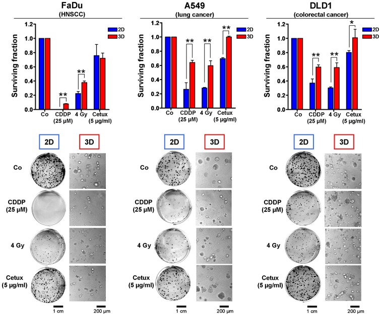
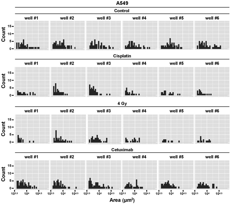
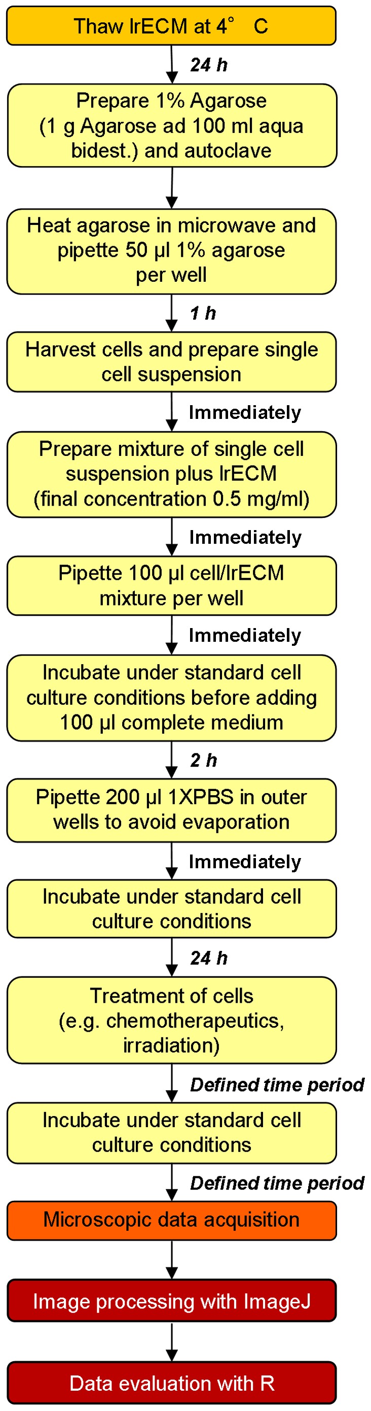
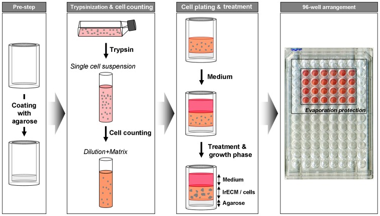
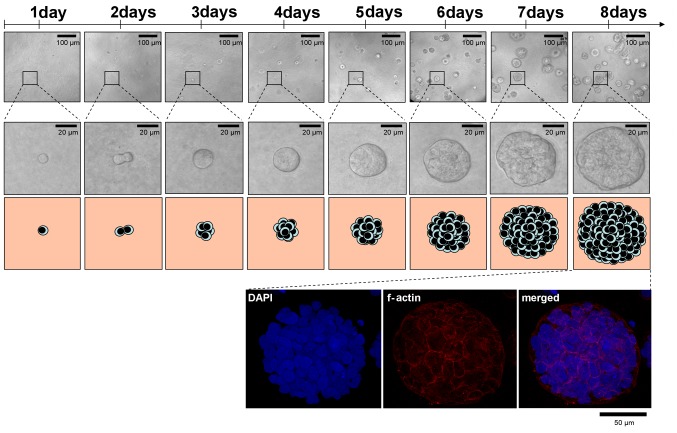
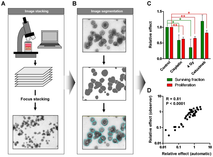
Three-dimensional ex vivo cell cultures mimic physiological in vivo growth conditions thereby significantly contributing to our understanding of tumor cell growth and survival, therapy resistance and identification of novel potent cancer targets. In the present study, we describe advanced three-dimensional cell culture methodology for investigating cellular survival and proliferation in human carcinoma cells after cancer therapy including molecular therapeutics. Single cells are embedded into laminin-rich extracellular matrix and can be treated with cytotoxic drugs, ionizing or UV radiation or any other substance of interest when consolidated and approximating in vivo morphology. Subsequently, cells are allowed to grow for automated determination of clonogenic survival (colony number) or proliferation (colony size). The entire protocol of 3D cell plating takes ~1 h working time and pursues for ~7 days before evaluation. This newly developed method broadens the spectrum of exploration of malignant tumors and other diseases and enables the obtainment of more reliable data on cancer treatment efficacy.
三维体外细胞培养模拟生理体内生长条件,从而极大地有助于我们理解肿瘤细胞的生长与存活、治疗抗性以及新型强效癌症靶点的识别。在本研究中,我们描述了先进的三维细胞培养方法,用于研究癌症治疗(包括分子疗法)后人类癌细胞的细胞存活和增殖情况。将单细胞嵌入富含层粘连蛋白的细胞外基质中,当细胞聚集并接近体内形态时,可用细胞毒性药物、电离辐射或紫外线辐射或任何其他感兴趣的物质进行处理。随后,让细胞生长,以便自动测定克隆形成存活率(集落数)或增殖情况(集落大小)。三维细胞铺板的整个流程需要约1小时的工作时间,在评估前持续约7天。这种新开发的方法拓宽了对恶性肿瘤和其他疾病的探索范围,并能够获得关于癌症治疗效果的更可靠数据。