Zhong Xiao-Mei, Zhang Fang, Yang Ming, Wen Xue-Hua, Zhang Xiang, Duan Xiao-Hui, Shen Jun
Department of Radiology, Sun Yat-Sen Memorial Hospital, Sun Yat-Sen University, No. 107 Yanjiang Road West, Guangzhou, Guangdong 510120, China.
Department of Radiology, Zhongda Hospital of Southeast University, Nanjing 210009, China.
Biomed Res Int. 2015;2015:131054. doi: 10.1155/2015/131054. Epub 2015 Oct 25.
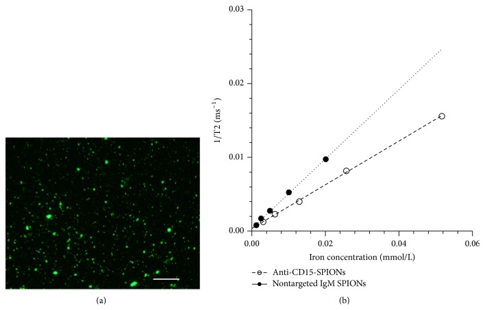
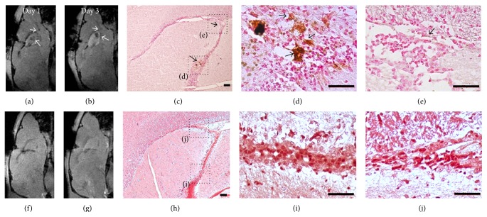
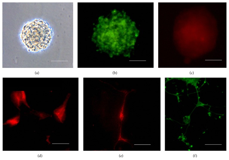
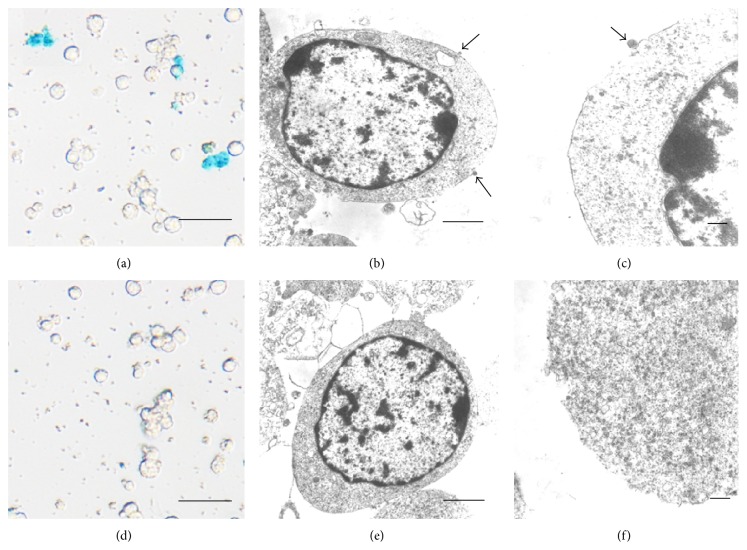
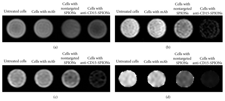
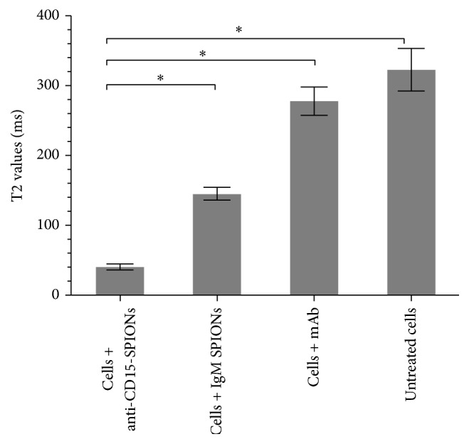
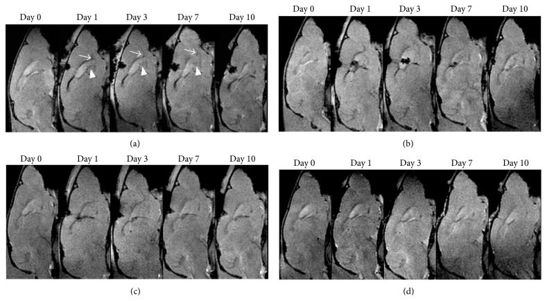
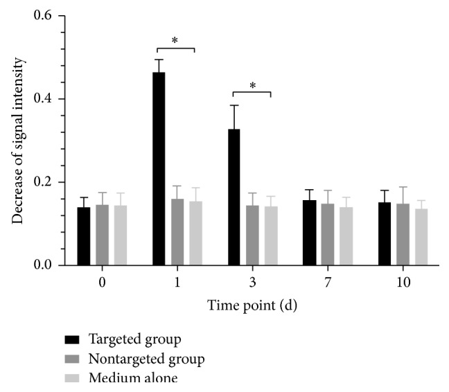
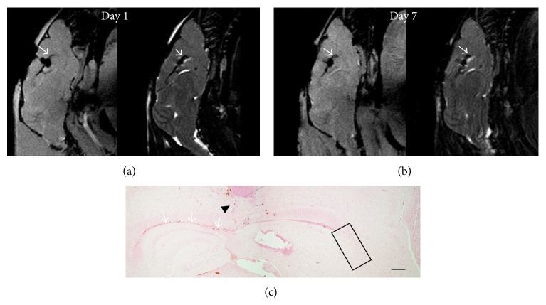
Neural stem cells in the adult mammalian brain have a significant level of neurogenesis plasticity. In vivo monitoring of adult endogenous NSCs would be of great benefit to the understanding of the neurogenesis plasticity under normal and pathological conditions. Here we show the feasibility of in vivo targeted MR imaging of endogenous NSCs in adult mouse brain by intraventricular delivery of monoclonal anti-CD15 antibody conjugated superparamagnetic iron oxide nanoparticles. After intraventricular administration of these nanoparticles, the subpopulation of NSCs in the anterior subventricular zone and the beginning of the rostral migratory stream could be in situ labeled and were in vivo visualized with 7.0-T MR imaging during a period from 1 day to 7 days after the injection. Histology confirmed that the injected targeted nanoparticles were specifically bound to CD15 positive cells and their surrounding extracellular matrix. Our results suggest that in vivo targeted MR imaging of endogenous neural stem cells in adult rodent brain could be achieved by using anti-CD15-SPIONs as the molecular probe; and this targeting imaging strategy has the advantage of a rapid in vivo monitoring of the subpopulation of endogenous NSCs in adult brains.
成年哺乳动物大脑中的神经干细胞具有显著水平的神经发生可塑性。对成年内源性神经干细胞进行体内监测将极大有助于理解正常和病理条件下的神经发生可塑性。在此,我们展示了通过脑室内递送单克隆抗CD15抗体偶联的超顺磁性氧化铁纳米颗粒,对成年小鼠脑内源性神经干细胞进行体内靶向磁共振成像的可行性。在脑室内给予这些纳米颗粒后,脑室下区前部和吻侧迁移流起始部位的神经干细胞亚群能够被原位标记,并在注射后1天至7天的时间段内通过7.0-T磁共振成像在体内可视化。组织学证实,注射的靶向纳米颗粒特异性结合到CD15阳性细胞及其周围的细胞外基质。我们的结果表明,使用抗CD15-超顺磁性氧化铁纳米颗粒作为分子探针可实现成年啮齿动物脑内源性神经干细胞的体内靶向磁共振成像;并且这种靶向成像策略具有能够快速在体内监测成年脑内源性神经干细胞亚群的优势。