Kong Wen-Tao, Ji Zheng-Biao, Wang Wen-Ping, Cai Hao, Huang Bei-Jian, Ding Hong
Department of Ultrasound, Zhongshan Hospital, Fudan University, Shanghai, China.
Gut Liver. 2016 Mar;10(2):283-7. doi: 10.5009/gnl14324.
BACKGROUND/AIMS: To evaluate the enhancement patterns of liver metastases and their influencing factors using dynamic contrast-enhanced ultrasound (CEUS).
A total of 240 patients (139 male and 101 female; 58.5 ± 11.2 years of age) diagnosed with liver metastases in our hospital were enrolled in this study to evaluate tumor characteristics using CEUS. A comparison of enhancement patterns with tumor size and primary tumor type was performed using the chi-square test. The differences between quantitative variables were evaluated with the independent-sample t-test and one-way analysis of variance.
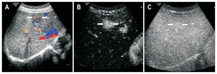
The enhancement patterns of liver metastases on CEUS were categorized as diffuse homogeneous hyperenhancement (133/240, 55.4%), rim-like hyperenhancement (80/240, 33.3%), heterogeneous hyperenhancement (10/240, 4.2%), and isoenhancement (17/240, 7.1%). There were significant differences in the enhancement patterns during the arterial phase based on the nodule size (p=0.001). A total of 231 of the nodules showed complete washout during the portal phase, and 237 nodules were hypoenhanced during the delayed phase. The washout time was correlated with tumor vascularity, with a longer washout time observed in hypervascular metastases compared to hypovascular metastases (p=0.033).
Diffuse homogeneous hyperenhancement followed by rapid washout was the most common enhancement pattern of liver metastases on CEUS and was affected by the nodule size and tumor vascularity. Small metastases were prone to show diffuse homogeneous hyperenhancement. Hypervascular metastases showed a significantly longer washout time compared to hypovascular metastases.
背景/目的:使用动态对比增强超声(CEUS)评估肝转移瘤的增强模式及其影响因素。
本研究纳入我院诊断为肝转移瘤的240例患者(男性139例,女性101例;年龄58.5±11.2岁),采用CEUS评估肿瘤特征。采用卡方检验比较增强模式与肿瘤大小和原发肿瘤类型。定量变量之间的差异采用独立样本t检验和单因素方差分析进行评估。
CEUS上肝转移瘤的增强模式分为弥漫性均匀高增强(133/240,55.4%)、环状高增强(80/240,33.3%)、不均匀高增强(10/240,4.2%)和等增强(17/240,7.1%)。基于结节大小,动脉期的增强模式存在显著差异(p=0.001)。共有231个结节在门静脉期表现为完全消退,237个结节在延迟期表现为低增强。消退时间与肿瘤血管有关,与乏血供转移瘤相比,富血供转移瘤的消退时间更长(p=0.033)。
弥漫性均匀高增强后快速消退是CEUS上肝转移瘤最常见的增强模式,且受结节大小和肿瘤血管影响。小转移瘤易表现为弥漫性均匀高增强。与乏血供转移瘤相比,富血供转移瘤的消退时间明显更长。