• 文献检索
  • 文档翻译
  • 深度研究
  • 学术资讯
  • Suppr Zotero 插件Zotero 插件
  • 邀请有礼
  • 套餐&价格
  • 历史记录
应用&插件
Suppr Zotero 插件Zotero 插件浏览器插件Mac 客户端Windows 客户端微信小程序
定价
高级版会员购买积分包购买API积分包
服务
文献检索文档翻译深度研究API 文档MCP 服务
关于我们
关于 Suppr公司介绍联系我们用户协议隐私条款
关注我们

Suppr 超能文献

核心技术专利:CN118964589B侵权必究
粤ICP备2023148730 号-1Suppr @ 2026

文献检索

告别复杂PubMed语法,用中文像聊天一样搜索,搜遍4000万医学文献。AI智能推荐,让科研检索更轻松。

立即免费搜索

文件翻译

保留排版,准确专业,支持PDF/Word/PPT等文件格式,支持 12+语言互译。

免费翻译文档

深度研究

AI帮你快速写综述,25分钟生成高质量综述,智能提取关键信息,辅助科研写作。

立即免费体验

基于显著性的3D卷积神经网络用于对多序列MRI上的常见肝脏局灶性病变进行分类

Saliency-based 3D convolutional neural network for categorising common focal liver lesions on multisequence MRI.

作者信息

Wang Shu-Hui, Han Xin-Jun, Du Jing, Wang Zhen-Chang, Yuan Chunwang, Chen Yinan, Zhu Yajing, Dou Xin, Xu Xiao-Wei, Xu Hui, Yang Zheng-Han

机构信息

Department of Radiology, Beijing Friendship Hospital, Capital Medical University, No. 95 Yongan Road, Xicheng District, Beijing, 100050, People's Republic of China.

Department of Radiology, Weihai Municipal Hospital, Cheeloo College of Medicine, Shandong University, Weihai, Shandong Province, People's Republic of China.

出版信息

Insights Imaging. 2021 Nov 24;12(1):173. doi: 10.1186/s13244-021-01117-z.

DOI:10.1186/s13244-021-01117-z
PMID:34817732
原文链接:https://pmc.ncbi.nlm.nih.gov/articles/PMC8613326/
Abstract

BACKGROUND

The imaging features of focal liver lesions (FLLs) are diverse and complex. Diagnosing FLLs with imaging alone remains challenging. We developed and validated an interpretable deep learning model for the classification of seven categories of FLLs on multisequence MRI and compared the differential diagnosis between the proposed model and radiologists.

METHODS

In all, 557 lesions examined by multisequence MRI were utilised in this retrospective study and divided into training-validation (n = 444) and test (n = 113) datasets. The area under the receiver operating characteristic curve (AUC) was calculated to evaluate the performance of the model. The accuracy and confusion matrix of the model and individual radiologists were compared. Saliency maps were generated to highlight the activation region based on the model perspective.

RESULTS

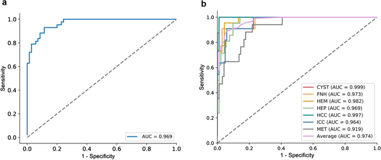
The AUC of the two- and seven-way classifications of the model were 0.969 (95% CI 0.944-0.994) and from 0.919 (95% CI 0.857-0.980) to 0.999 (95% CI 0.996-1.000), respectively. The model accuracy (79.6%) of the seven-way classification was higher than that of the radiology residents (66.4%, p = 0.035) and general radiologists (73.5%, p = 0.346) but lower than that of the academic radiologists (85.4%, p = 0.291). Confusion matrices showed the sources of diagnostic errors for the model and individual radiologists for each disease. Saliency maps detected the activation regions associated with each predicted class.

CONCLUSION

This interpretable deep learning model showed high diagnostic performance in the differentiation of FLLs on multisequence MRI. The analysis principle contributing to the predictions can be explained via saliency maps.

摘要

背景

肝脏局灶性病变(FLLs)的影像学特征多样且复杂。仅依靠影像学诊断FLLs仍具有挑战性。我们开发并验证了一种可解释的深度学习模型,用于在多序列磁共振成像(MRI)上对七类FLLs进行分类,并比较了该模型与放射科医生之间的鉴别诊断能力。

方法

本回顾性研究共纳入了557个经多序列MRI检查的病变,并将其分为训练-验证集(n = 444)和测试集(n = 113)。计算受试者操作特征曲线(ROC)下的面积(AUC)以评估模型的性能。比较了模型和个体放射科医生的准确性及混淆矩阵。基于模型视角生成显著性图以突出激活区域。

结果

该模型的二元和七元分类的AUC分别为0.969(95%可信区间0.944 - 0.994)和0.919(95%可信区间0.857 - 0.980)至0.999(95%可信区间0.996 - 1.000)。七元分类的模型准确率(79.6%)高于放射科住院医师(66.4%,p = 0.035)和普通放射科医生(73.5%,p = 0.346),但低于学术放射科医生(85.4%,p = 0.291)。混淆矩阵显示了模型和个体放射科医生对每种疾病的诊断错误来源。显著性图检测到了与每个预测类别相关的激活区域。

结论

这种可解释的深度学习模型在多序列MRI上对FLLs的鉴别诊断中表现出较高的诊断性能。有助于预测的分析原理可通过显著性图进行解释。

https://cdn.ncbi.nlm.nih.gov/pmc/blobs/9f5e/8613326/ea1fc0091efc/13244_2021_1117_Fig2_HTML.jpg

相似文献

1
Saliency-based 3D convolutional neural network for categorising common focal liver lesions on multisequence MRI.基于显著性的3D卷积神经网络用于对多序列MRI上的常见肝脏局灶性病变进行分类
Insights Imaging. 2021 Nov 24;12(1):173. doi: 10.1186/s13244-021-01117-z.
2
Multiphase convolutional dense network for the classification of focal liver lesions on dynamic contrast-enhanced computed tomography.基于动态对比增强 CT 的多相卷积密集网络的肝脏局灶性病变分类。
World J Gastroenterol. 2020 Jul 7;26(25):3660-3672. doi: 10.3748/wjg.v26.i25.3660.
3
Improving B-mode ultrasound diagnostic performance for focal liver lesions using deep learning: A multicentre study.利用深度学习提高局灶性肝病变的 B 型超声诊断性能:一项多中心研究。
EBioMedicine. 2020 Jun;56:102777. doi: 10.1016/j.ebiom.2020.102777. Epub 2020 Apr 28.
4
Deep learning radiomics for focal liver lesions diagnosis on long-range contrast-enhanced ultrasound and clinical factors.基于远程对比增强超声和临床因素的深度学习放射组学在肝脏局灶性病变诊断中的应用
Quant Imaging Med Surg. 2022 Jun;12(6):3213-3226. doi: 10.21037/qims-21-1004.
5
A deep dive into understanding tumor foci classification using multiparametric MRI based on convolutional neural network.基于卷积神经网络,深入探究利用多参数磁共振成像进行肿瘤病灶分类。
Med Phys. 2020 Sep;47(9):4077-4086. doi: 10.1002/mp.14255. Epub 2020 Jun 12.
6
Deep-learning-assisted diagnosis for knee magnetic resonance imaging: Development and retrospective validation of MRNet.深度学习辅助膝关节磁共振成像诊断:MRNet 的开发和回顾性验证。
PLoS Med. 2018 Nov 27;15(11):e1002699. doi: 10.1371/journal.pmed.1002699. eCollection 2018 Nov.
7
Deep learning for liver tumor diagnosis part II: convolutional neural network interpretation using radiologic imaging features.深度学习在肝脏肿瘤诊断中的应用 Ⅱ:利用影像学特征进行卷积神经网络解释。
Eur Radiol. 2019 Jul;29(7):3348-3357. doi: 10.1007/s00330-019-06214-8. Epub 2019 May 15.
8
Deep learning for liver tumor diagnosis part I: development of a convolutional neural network classifier for multi-phasic MRI.深度学习在肝脏肿瘤诊断中的应用 第一部分:用于多期 MRI 的卷积神经网络分类器的开发。
Eur Radiol. 2019 Jul;29(7):3338-3347. doi: 10.1007/s00330-019-06205-9. Epub 2019 Apr 23.
9
Diagnosis of focal liver lesions with deep learning-based multi-channel analysis of hepatocyte-specific contrast-enhanced magnetic resonance imaging.基于深度学习的肝细胞特异性对比增强磁共振成像多通道分析对肝脏局灶性病变的诊断。
World J Gastroenterol. 2021 Sep 21;27(35):5978-5988. doi: 10.3748/wjg.v27.i35.5978.
10
Attention-based Saliency Maps Improve Interpretability of Pneumothorax Classification.基于注意力的显著图提高气胸分类的可解释性。
Radiol Artif Intell. 2022 Mar 1;5(2):e220187. doi: 10.1148/ryai.220187. eCollection 2023 Mar.

引用本文的文献

1
Application of biological big data and radiomics in hepatocellular carcinoma.生物大数据和放射组学在肝细胞癌中的应用。
ILIVER. 2023 Feb 4;2(1):41-49. doi: 10.1016/j.iliver.2023.01.003. eCollection 2023 Mar.
2
A two-step automatic identification of contrast phases for abdominal CT images based on residual networks.基于残差网络的腹部CT图像对比剂阶段两步自动识别
Insights Imaging. 2025 Jun 27;16(1):139. doi: 10.1186/s13244-025-01995-7.
3
Evaluating artificial intelligence for a focal nodular hyperplasia diagnosis using magnetic resonance imaging: preliminary findings.

本文引用的文献

1
Automatic Detection and Classification of Focal Liver Lesions Based on Deep Convolutional Neural Networks: A Preliminary Study.基于深度卷积神经网络的肝脏局灶性病变自动检测与分类:一项初步研究。
Front Oncol. 2021 Jan 29;10:581210. doi: 10.3389/fonc.2020.581210. eCollection 2020.
2
Deep Learning for Accurate Diagnosis of Liver Tumor Based on Magnetic Resonance Imaging and Clinical Data.基于磁共振成像和临床数据的深度学习用于肝脏肿瘤的准确诊断
Front Oncol. 2020 May 28;10:680. doi: 10.3389/fonc.2020.00680. eCollection 2020.
3
Assessing Radiology Research on Artificial Intelligence: A Brief Guide for Authors, Reviewers, and Readers-From the Editorial Board.
利用磁共振成像评估人工智能用于局灶性结节性增生诊断:初步结果。
Diagn Interv Radiol. 2025 Mar 26. doi: 10.4274/dir.2025.243095.
4
SenseCare: a research platform for medical image informatics and interactive 3D visualization.SenseCare:一个用于医学图像信息学和交互式3D可视化的研究平台。
Front Radiol. 2024 Nov 21;4:1460889. doi: 10.3389/fradi.2024.1460889. eCollection 2024.
5
Automatic localization and deep convolutional generative adversarial network-based classification of focal liver lesions in computed tomography images: A preliminary study.基于自动定位和深度卷积生成对抗网络的计算机断层扫描图像中肝脏局灶性病变分类:一项初步研究。
J Gastroenterol Hepatol. 2025 Jan;40(1):166-176. doi: 10.1111/jgh.16803. Epub 2024 Nov 14.
6
Deep learning model based on contrast-enhanced MRI for predicting post-surgical survival in patients with hepatocellular carcinoma.基于对比增强磁共振成像的深度学习模型用于预测肝细胞癌患者术后生存率
Heliyon. 2024 May 16;10(11):e31451. doi: 10.1016/j.heliyon.2024.e31451. eCollection 2024 Jun 15.
7
Deep Learning Methods in Medical Image-Based Hepatocellular Carcinoma Diagnosis: A Systematic Review and Meta-Analysis.基于医学图像的肝细胞癌诊断中的深度学习方法:系统评价与荟萃分析
Cancers (Basel). 2023 Dec 3;15(23):5701. doi: 10.3390/cancers15235701.
8
MAIC-10 brief quality checklist for publications using artificial intelligence and medical images.用于使用人工智能和医学图像的出版物的MAIC-10简要质量检查表。
Insights Imaging. 2023 Jan 16;14(1):11. doi: 10.1186/s13244-022-01355-9.
9
Focal Liver Lesion MRI Feature Identification Using Efficientnet and MONAI: A Feasibility Study.基于 Efficientnet 和 MONAI 的肝脏局灶性病变 MRI 特征识别:一项可行性研究。
Cells. 2022 May 5;11(9):1558. doi: 10.3390/cells11091558.
评估人工智能放射学研究:给作者、审稿人和读者的简要指南——来自编辑委员会
Radiology. 2020 Mar;294(3):487-489. doi: 10.1148/radiol.2019192515. Epub 2019 Dec 31.
4
Non-invasive diagnostic criteria of hepatocellular carcinoma: Comparison of diagnostic accuracy of updated LI-RADS with clinical practice guidelines of OPTN-UNOS, AASLD, NCCN, EASL-EORTC, and KLSCG-NCC.肝细胞癌的无创性诊断标准:LI-RADS 更新版与 OPTN-UNOS、AASLD、NCCN、EASL-EORTC 和 KLSCG-NCC 临床实践指南的诊断准确性比较。
PLoS One. 2019 Dec 10;14(12):e0226291. doi: 10.1371/journal.pone.0226291. eCollection 2019.
5
Medical Image Perception Research in the Emerging Age of Artificial Intelligence.
Radiology. 2020 Jan;294(1):210-211. doi: 10.1148/radiol.2019192252. Epub 2019 Nov 12.
6
Artificial intelligence in medical imaging of the liver.人工智能在肝脏医学影像中的应用。
World J Gastroenterol. 2019 Feb 14;25(6):672-682. doi: 10.3748/wjg.v25.i6.672.
7
Medical Image Analysis using Convolutional Neural Networks: A Review.基于卷积神经网络的医学图像分析:综述
J Med Syst. 2018 Oct 8;42(11):226. doi: 10.1007/s10916-018-1088-1.
8
Validation of Liver Imaging Reporting and Data System 2017 (LI-RADS) Criteria for Imaging Diagnosis of Hepatocellular Carcinoma.验证 2017 版肝脏影像报告和数据系统(LI-RADS)标准在肝细胞癌影像学诊断中的应用。
J Magn Reson Imaging. 2019 Jun;49(7):e205-e215. doi: 10.1002/jmri.26329. Epub 2018 Sep 26.
9
Deep dense multi-path neural network for prostate segmentation in magnetic resonance imaging.深度密集多路径神经网络在磁共振成像中的前列腺分割。
Int J Comput Assist Radiol Surg. 2018 Nov;13(11):1687-1696. doi: 10.1007/s11548-018-1841-4. Epub 2018 Aug 7.
10
Artificial intelligence in radiology: how will we be affected?放射学中的人工智能:我们将受到怎样的影响?
Eur Radiol. 2019 Jan;29(1):141-143. doi: 10.1007/s00330-018-5644-3. Epub 2018 Jul 19.