Krämer Johannes, Bijnens Bart, Störk Stefan, Ritter Christian O, Liu Dan, Ertl Georg, Wanner Christoph, Weidemann Frank
Department of Medicine I, Cardiology Unit, University of Würzburg, Würzburg, Germany.
Department of Pediatrics and Adolescent Medicine, University of Ulm, Ulm, Germany.
PLoS One. 2015 Nov 23;10(11):e0140627. doi: 10.1371/journal.pone.0140627. eCollection 2015.
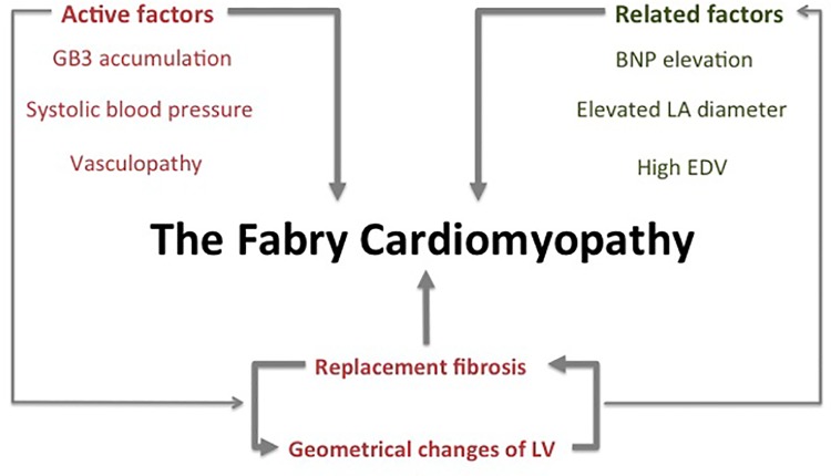
In spite of several research studies help to describe the heart in Fabry disease (FD), the cardiomyopathy is not entirely understood. In addition, the impact of blood pressure and alterations in geometry have not been systematically evaluated.
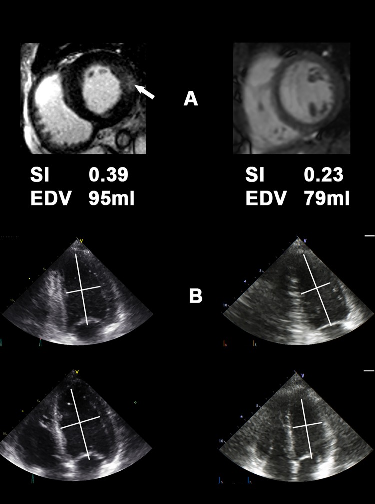
In 74 FD patients (mean age 36±12 years; 45 females) the extent of myocardial fibrosis and its progression were quantified using cardiac magnetic-resonance-imaging with late enhancement technique (LE). Results were compared to standard echocardiography complemented by 2D-speckle-tracking, 3D-sphericity-index (SI) and standardized blood pressure measurement. At baseline, no patient received enzyme replacement therapy (ERT). After 51±24 months, a follow-up examination was performed.
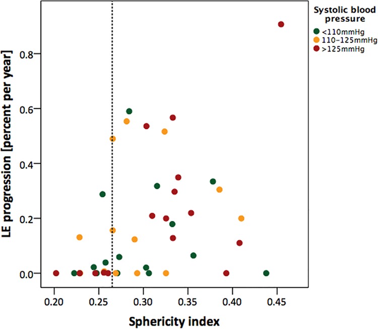
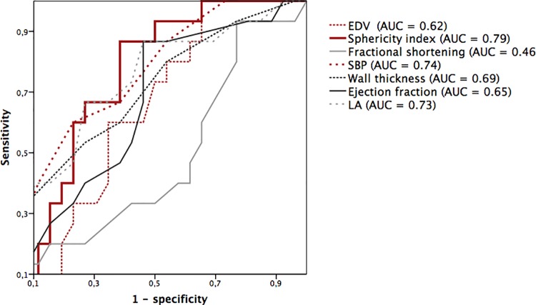
Systolic blood pressure (SBP) was higher in patients with vs. without LE: 123±17 mmHg vs. 115±13 mmHg; P = 0.04. A positive correlation was found between SI and the amount of LE-positive myocardium (r = 0.51; P<0.001) indicating an association of higher SI in more advanced stages of the cardiomyopathy. SI at baseline was positively associated with the increase of LE-positive myocardium during follow-up. The highest SBP (125±19 mmHg) and also the highest SI (0.32±0.05) was found in the subgroup with a rapidly increasing LE (ie, ≥0.2% per year; n = 16; P = 0.04). Multivariate logistic regression analysis including SI, SBP, EF, left ventricular volumes, wall thickness and NT-proBNP adjusted for age and sex showed SI as the most powerful parameter to detect rapid progression of LE (AUC = 0.785; P<0.05).
LV geometry as assessed by the sphericity index is altered in relation to the stage of the Fabry cardiomyopathy. Although patients with FD are not hypertensive, the SBP has a clear impact on the progression of the cardiomyopathy.
尽管有多项研究有助于描述法布里病(FD)患者的心脏情况,但对其心肌病仍未完全了解。此外,血压和几何形态改变的影响尚未得到系统评估。
在74例FD患者(平均年龄36±12岁;45例女性)中,采用心脏磁共振成像延迟强化技术(LE)对心肌纤维化程度及其进展进行量化。将结果与标准超声心动图(辅以二维斑点追踪、三维球形指数(SI)和标准化血压测量)进行比较。基线时,无患者接受酶替代疗法(ERT)。51±24个月后进行随访检查。
有LE的患者收缩压(SBP)高于无LE的患者:123±17 mmHg对115±13 mmHg;P = 0.04。SI与LE阳性心肌量之间存在正相关(r = 0.51;P<0.001),表明在心肌病更晚期阶段SI较高。基线时的SI与随访期间LE阳性心肌的增加呈正相关。在LE快速增加(即每年≥0.2%;n = 16;P = 0.04)的亚组中发现最高SBP(125±19 mmHg)和最高SI(0.32±0.05)。多因素逻辑回归分析纳入SI、SBP、EF、左心室容积、壁厚和NT-proBNP,并对年龄和性别进行校正,结果显示SI是检测LE快速进展的最有力参数(AUC = !0.785;P<0.05)。
根据球形指数评估的左心室几何形态与法布里心肌病的阶段相关。尽管FD患者并非高血压患者,但SBP对心肌病的进展有明显影响。