Soh Pauline, Narayanan Balaji, Khadka Sabin, Calhoun Vince D, Keshavan Matcheri S, Tamminga Carol A, Sweeney John A, Clementz Brett A, Pearlson Godfrey D
Olin Neuropsychiatry Research Center, Institute of Living , Hartford, CT , USA.
Department of Electrical and Computer Engineering, University of New Mexico , Albuquerque, NM , USA ; The Mind Research Network , Albuquerque, NM , USA ; Department of Psychiatry, Yale University School of Medicine , New Haven, CT , USA.
Front Psychiatry. 2015 Nov 9;6:162. doi: 10.3389/fpsyt.2015.00162. eCollection 2015.
Many studies have examined either electroencephalogram (EEG) frequency activity or gray matter volumes (GMV) in various psychoses [including schizophrenia (SZ), schizoaffective (SZA), and psychotic bipolar disorder (PBP)]. Prior work demonstrated similar EEG and gray matter abnormalities in both SZ and PBP. Integrating EEG and GMV and jointly analyzing the combined data fully elucidates the linkage between the two and may provide better biomarker- or endophenotype-specificity for a particular illness. Joint exploratory investigations of EEG and GMV are scarce in the literature and the relationship between the two in psychosis is even less explored. We investigated a joint multivariate model to test whether the linear relationship or linkage between awake EEG (AEEG) frequency activity and GMV is abnormal across the psychosis dimension and if such effects are also present in first-degree relatives.
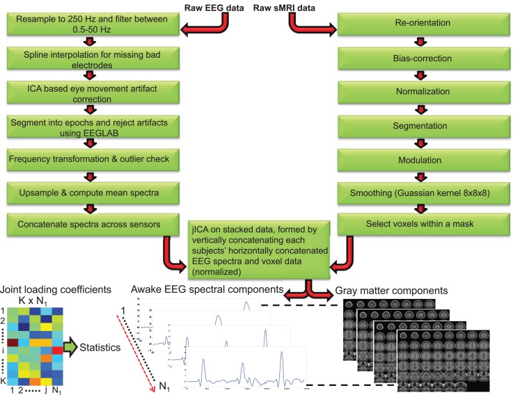
We assessed 607 subjects comprising 264 probands [105 SZ, 72 SZA, and 87 PBP], 233 of their first degree relatives [82 SZ relatives (SZR), 71 SZA relatives (SZAR), and 80 PBP relatives (PBPR)], and 110 healthy comparison subjects (HC). All subjects underwent structural MRI (sMRI) and EEG scans. Frequency activity and voxel-based morphometric GMV were derived from EEG and sMRI data, respectively. Seven AEEG frequency and gray matter components were extracted using Joint independent component analysis (jICA). The loading coefficients (LC) were examined for group differences using analysis of covariance. Further, the LCs were correlated with psychopathology scores to identify relationship with clinical symptoms.
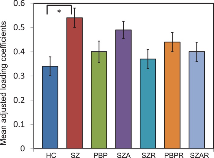
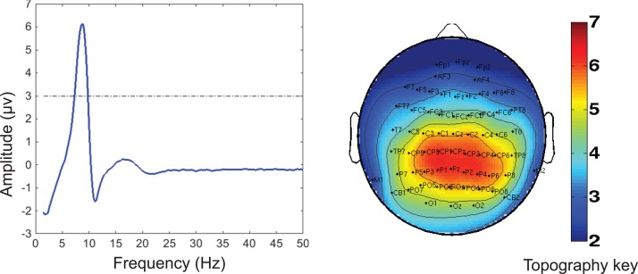
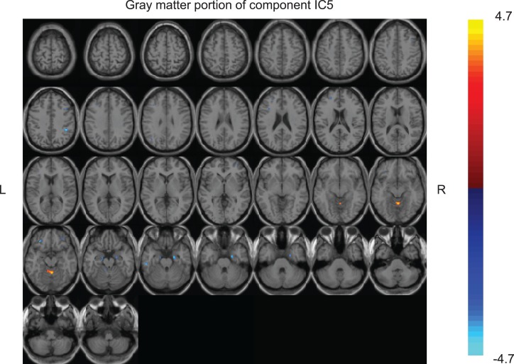
Joint ICA revealed a single component differentiating SZ from HC (p < 0.006), comprising increased posterior alpha activity associated with decreased volume in inferior parietal lobe, supramarginal, parahippocampal gyrus, middle frontal, inferior temporal gyri, and increased volume of uncus and culmen. No components were aberrant in either PBP or SZA or any relative group. No significant association was identified with clinical symptom measures.
Our data suggest that a joint EEG and GMV model yielded a biomarker specific to SZ, not abnormal in PBP or SZA. Alpha activity was related to both increased and decreased volume in different cortical structures. Additionally, the joint model failed to identify endophenotypes across psychotic disorders.
许多研究考察了各种精神病(包括精神分裂症(SZ)、分裂情感性障碍(SZA)和精神病性双相障碍(PBP))的脑电图(EEG)频率活动或灰质体积(GMV)。先前的研究表明,SZ和PBP在EEG和灰质方面均存在类似异常。整合EEG和GMV并联合分析综合数据,能够充分阐明两者之间的联系,并可能为特定疾病提供更好的生物标志物或内表型特异性。EEG和GMV的联合探索性研究在文献中较为少见,两者在精神病中的关系更是鲜有探讨。我们研究了一个联合多变量模型,以检验清醒EEG(AEEG)频率活动与GMV之间的线性关系或联系在整个精神病维度上是否异常,以及这种效应在一级亲属中是否也存在。
我们评估了607名受试者,包括264名先证者[105名SZ、72名SZA和87名PBP]、他们的233名一级亲属[82名SZ亲属(SZR)、71名SZA亲属(SZAR)和80名PBP亲属(PBPR)]以及110名健康对照受试者(HC)。所有受试者均接受了结构MRI(sMRI)和EEG扫描。频率活动和基于体素的形态学GMV分别从EEG和sMRI数据中得出。使用联合独立成分分析(jICA)提取七个AEEG频率和灰质成分。使用协方差分析检查负荷系数(LC)的组间差异。此外,将LC与精神病理学评分相关联,以确定与临床症状的关系。
联合ICA显示出一个区分SZ与HC的单一成分(p < 0.006),包括后α活动增加,同时顶下小叶、缘上回、海马旁回、额中回、颞下回体积减小,钩回和山顶体积增加。PBP、SZA或任何亲属组中均未发现异常成分。未发现与临床症状指标有显著关联。
我们的数据表明,EEG和GMV联合模型产生了一种特定于SZ的生物标志物,在PBP或SZA中无异常。α活动与不同皮质结构的体积增加和减小均有关。此外,联合模型未能识别出跨精神病性障碍的内表型。