Ferreira Bianca Lima, Orikaza Cristina Mary, Cordero Esteban Mauricio, Mortara Renato Arruda
Departamento de Microbiologia, Imunologia e Parasitologia, Escola Paulista de Medicina, Universidade Federal de São Paulo (EPM-UNIFESP), Rua Botucatu 862 6th floor, São Paulo, São Paulo, Brazil.
Cell Microbiol. 2016 Jun;18(6):779-83. doi: 10.1111/cmi.12553. Epub 2016 Jan 5.
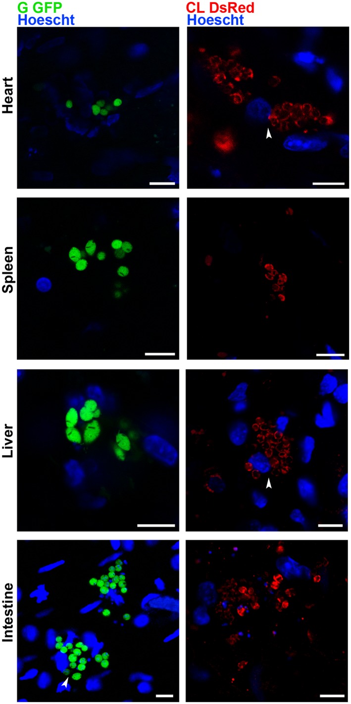
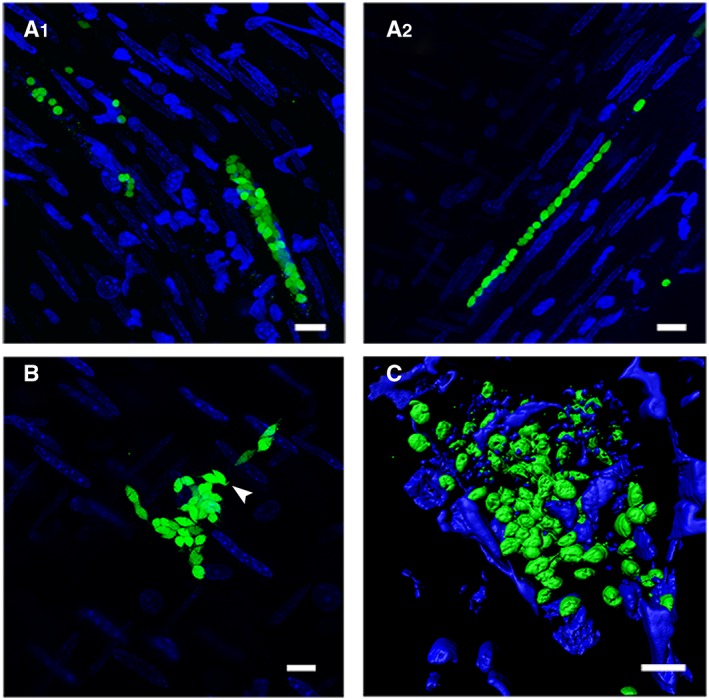
Although imaging the live Trypanosoma cruzi parasite is a routine technique in most laboratories, identification of the parasite in infected tissues and organs has been hindered by their intrinsic opaque nature. We describe a simple method for in vivo observation of live single-cell Trypanosoma cruzi parasites inside mammalian host tissues. BALB/c or C57BL/6 mice infected with DsRed-CL or GFP-G trypomastigotes had their organs removed and sectioned with surgical blades. Ex vivo organ sections were observed under confocal microscopy. For the first time, this procedure enabled imaging of individual amastigotes, intermediate forms and motile trypomastigotes within infected tissues of mammalian hosts.
尽管对活的克氏锥虫寄生虫进行成像在大多数实验室中是一项常规技术,但由于受感染组织和器官本身不透明的特性,在其中鉴定寄生虫一直受到阻碍。我们描述了一种在哺乳动物宿主组织内对活的单细胞克氏锥虫寄生虫进行体内观察的简单方法。用DsRed-CL或GFP-G型锥鞭毛体感染的BALB/c或C57BL/6小鼠的器官被切除并用手术刀片切片。在共聚焦显微镜下观察离体器官切片。首次通过该程序能够对哺乳动物宿主受感染组织内的单个无鞭毛体、中间形态和活动的锥鞭毛体进行成像。