Li Zuosheng, Li Xin, Song Zuoqing, Liu Jinghao, Dong Ming, Shi Tao, Ren Dian, Xu Song, Chen Jun
Department of Lung Cancer Surgery, Lung Cancer Institute, Tianjin Medical University General Hospital, No. 154 Anshan Road, Heping District, Tianjin, 300052, China.
Department of Thoracic Surgery, North China University of Science and Technology Affiliated Hospital, Tangshan, 063000, China.
World J Surg Oncol. 2015 Dec 12;13:333. doi: 10.1186/s12957-015-0748-6.
Sarcoidosis is a rare condition that is often misdiagnosed as malignant tumors due to the similar clinical manifestations and imaging findings.
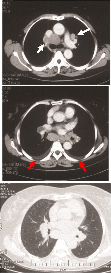
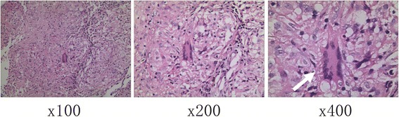
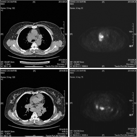
We encountered a 56-year-old Chinese woman who had a chief complaint of a persistent cough. The chest computer tomography (CT) revealed mediastinal and bilateral hilar lymph node enlargement, and positron emission tomography-computer tomography (PET-CT) revealed abnormal fluorodeoxyglucose (FDG) uptake in the lymph nodes of the chest and abdomen. To further clarify the diagnosis, a lymph node sampling was performed by video-assisted thoracoscopic surgery (VATS) and the histopathologic diagnosis of sarcoidosis was confirmed.
VATS could be an effective and minimally invasive diagnostic method to discriminate pulmonary sarcoidosis with other malignant tumors.
结节病是一种罕见疾病,常因临床表现和影像学表现相似而被误诊为恶性肿瘤。
我们接诊了一名56岁的中国女性,主要症状为持续咳嗽。胸部计算机断层扫描(CT)显示纵隔及双侧肺门淋巴结肿大,正电子发射断层扫描-计算机断层扫描(PET-CT)显示胸部和腹部淋巴结氟脱氧葡萄糖(FDG)摄取异常。为进一步明确诊断,通过电视辅助胸腔镜手术(VATS)进行了淋巴结采样,确诊为结节病。
VATS可能是鉴别肺结节病与其他恶性肿瘤的一种有效且微创的诊断方法。