• 文献检索
  • 文档翻译
  • 深度研究
  • 学术资讯
  • Suppr Zotero 插件Zotero 插件
  • 邀请有礼
  • 套餐&价格
  • 历史记录
应用&插件
Suppr Zotero 插件Zotero 插件浏览器插件Mac 客户端Windows 客户端微信小程序
定价
高级版会员购买积分包购买API积分包
服务
文献检索文档翻译深度研究API 文档MCP 服务
关于我们
关于 Suppr公司介绍联系我们用户协议隐私条款
关注我们

Suppr 超能文献

核心技术专利:CN118964589B侵权必究
粤ICP备2023148730 号-1Suppr @ 2026

文献检索

告别复杂PubMed语法,用中文像聊天一样搜索,搜遍4000万医学文献。AI智能推荐,让科研检索更轻松。

立即免费搜索

文件翻译

保留排版,准确专业,支持PDF/Word/PPT等文件格式,支持 12+语言互译。

免费翻译文档

深度研究

AI帮你快速写综述,25分钟生成高质量综述,智能提取关键信息,辅助科研写作。

立即免费体验

波兰超声学会标准 - 更新。脾脏检查。

Standards of the Polish Ultrasound Society - update. Spleen examination.

作者信息

Walczyk Joanna, Walas Maria Krystyna

机构信息

Oddział Kliniczny Kliniki Chorób Metabolicznych, Szpital Uniwersytecki, Kraków, Polska.

出版信息

J Ultrason. 2013 Mar;13(52):50-64. doi: 10.15557/JoU.2013.0004. Epub 2013 Mar 30.

DOI:10.15557/JoU.2013.0004
PMID:26672802
原文链接:https://pmc.ncbi.nlm.nih.gov/articles/PMC4613573/
Abstract

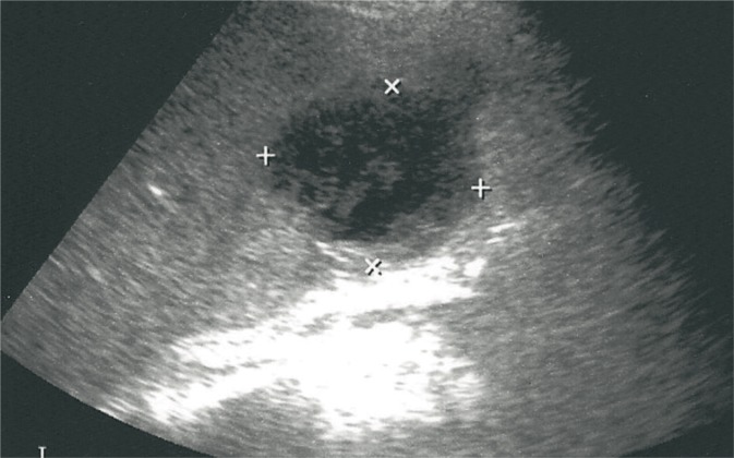
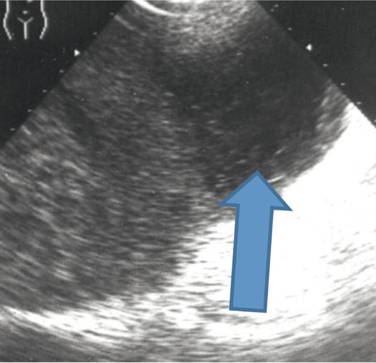
Ultrasound scan of the spleen is an integral part of the overall abdominal examination. Due to its anatomical position, physical examination of the spleen is frequently supplemented with an ultrasound which plays a special role in the differential diagnostics of splenic diseases and facilitates the determination of further diagnostic and therapeutic procedures. Similarly to other types of ultrasound scans, the examiner should be familiar with all significant clinical information as well as results of examinations and tests conducted so far. This enables to narrow the scope of search for etiological factors and indicate specific disease entities in the findings as well as allows for accurate assessment of coexistent pathologies. The article presents the standards of the Polish Ultrasound Society concerning the apparatus, preparation for the examination, technique and description of the findings. The authors discuss the normal anatomy of the spleen and the most common pathologies ranging from splenomegaly to splenic traumas. The indications for the contrast-enhanced ultrasound and characteristic patterns of enhancement of individual focal lesions are presented. This article is supplemented with photographic documentation, which provides images of the discussed lesions. The ultrasound examination, if carried out in compliance with current standards, allows for accurate interpretation of detected changes. This article has been prepared on the basis of the Ultrasound Examination Standards of the Polish Ultrasound Society (2011) and updated with the current knowledge.

摘要

脾脏超声检查是整个腹部检查不可或缺的一部分。由于脾脏的解剖位置,脾脏的体格检查常常需要辅以超声检查,超声在脾脏疾病的鉴别诊断中发挥着特殊作用,并有助于确定进一步的诊断和治疗程序。与其他类型的超声检查一样,检查者应熟悉所有重要的临床信息以及迄今为止进行的检查和测试结果。这有助于缩小对病因的搜索范围,在检查结果中指出具体的疾病实体,并能准确评估并存的病理情况。本文介绍了波兰超声学会关于仪器设备、检查准备、技术及检查结果描述的标准。作者讨论了脾脏的正常解剖结构以及从脾肿大到脾外伤等最常见的病理情况。文中还讨论了超声造影的适应证以及各个局灶性病变的特征性增强模式。本文配有照片资料,展示了所讨论病变的图像。如果按照现行标准进行超声检查,就能对检测到的变化进行准确解读。本文是根据波兰超声学会《超声检查标准》(2011年)编写,并结合当前知识进行了更新。

https://cdn.ncbi.nlm.nih.gov/pmc/blobs/54e3/4613573/9e010b26cdee/JoU-2013-0004-g017.jpg
https://cdn.ncbi.nlm.nih.gov/pmc/blobs/54e3/4613573/65d358360dd3/JoU-2013-0004-g001.jpg
https://cdn.ncbi.nlm.nih.gov/pmc/blobs/54e3/4613573/6fe466a34bf8/JoU-2013-0004-g002.jpg
https://cdn.ncbi.nlm.nih.gov/pmc/blobs/54e3/4613573/71715e1294cb/JoU-2013-0004-g003.jpg
https://cdn.ncbi.nlm.nih.gov/pmc/blobs/54e3/4613573/04e709009cf5/JoU-2013-0004-g004.jpg
https://cdn.ncbi.nlm.nih.gov/pmc/blobs/54e3/4613573/fcec66bbe01c/JoU-2013-0004-g005.jpg
https://cdn.ncbi.nlm.nih.gov/pmc/blobs/54e3/4613573/0acfdb2d9c45/JoU-2013-0004-g006.jpg
https://cdn.ncbi.nlm.nih.gov/pmc/blobs/54e3/4613573/ea9082bbe1bf/JoU-2013-0004-g007.jpg
https://cdn.ncbi.nlm.nih.gov/pmc/blobs/54e3/4613573/1671440f26c4/JoU-2013-0004-g008.jpg
https://cdn.ncbi.nlm.nih.gov/pmc/blobs/54e3/4613573/656d1e01906b/JoU-2013-0004-g009.jpg
https://cdn.ncbi.nlm.nih.gov/pmc/blobs/54e3/4613573/fd0ba3f81185/JoU-2013-0004-g010.jpg
https://cdn.ncbi.nlm.nih.gov/pmc/blobs/54e3/4613573/1a025ae26ba7/JoU-2013-0004-g011.jpg
https://cdn.ncbi.nlm.nih.gov/pmc/blobs/54e3/4613573/343c5b1e4e99/JoU-2013-0004-g012.jpg
https://cdn.ncbi.nlm.nih.gov/pmc/blobs/54e3/4613573/98b790fe2eb8/JoU-2013-0004-g013.jpg
https://cdn.ncbi.nlm.nih.gov/pmc/blobs/54e3/4613573/4c3363b7e7ab/JoU-2013-0004-g014.jpg
https://cdn.ncbi.nlm.nih.gov/pmc/blobs/54e3/4613573/8e6926e4fa9d/JoU-2013-0004-g015.jpg
https://cdn.ncbi.nlm.nih.gov/pmc/blobs/54e3/4613573/8cdf02946829/JoU-2013-0004-g016.jpg
https://cdn.ncbi.nlm.nih.gov/pmc/blobs/54e3/4613573/9e010b26cdee/JoU-2013-0004-g017.jpg

相似文献

1
Standards of the Polish Ultrasound Society - update. Spleen examination.波兰超声学会标准 - 更新。脾脏检查。
J Ultrason. 2013 Mar;13(52):50-64. doi: 10.15557/JoU.2013.0004. Epub 2013 Mar 30.
2
Errors made in the ultrasound diagnostics of the spleen.脾脏超声诊断中的错误。
J Ultrason. 2013 Mar;13(52):65-72. doi: 10.15557/JoU.2013.0005. Epub 2013 Mar 30.
3
Standards of the Polish Ultrasound Society - update. The liver, gallbladder and bile ducts examinations.波兰超声学会标准——更新。肝脏、胆囊及胆管检查。
J Ultrason. 2012 Dec;12(51):428-45. doi: 10.15557/JoU.2012.0031. Epub 2012 Dec 30.
4
Standards of the Polish Ultrasound Society - update. Pancreas examination.波兰超声学会标准 - 更新。胰腺检查。
J Ultrason. 2013 Jun;13(53):167-77. doi: 10.15557/JoU.2013.0017. Epub 2013 Jun 30.
5
Standards of the Polish Ultrasound Society - update. Sonomammography examination.波兰超声学会标准——更新。乳腺超声检查。
J Ultrason. 2012 Sep;12(50):245-61. doi: 10.15557/JoU.2012.0010. Epub 2012 Sep 30.
6
Standards of the Polish Ultrasound Society - update. Ultrasound examination of thyroid gland and ultrasound-guided thyroid biopsy.波兰超声学会标准——更新。甲状腺超声检查及超声引导下甲状腺活检。
J Ultrason. 2014 Mar;14(56):49-60. doi: 10.15557/JoU.2014.0005. Epub 2014 Mar 30.
7
Real-time contrast-enhanced ultrasound of the spleen: examination technique and preliminary clinical experience.脾脏实时超声造影:检查技术及初步临床经验
Radiol Med. 2003 Oct;106(4):338-56.
8
Diagnostic insights into splenic pathologies: the role of multiparametric ultrasound.脾脏疾病的诊断见解:多参数超声的作用
Abdom Radiol (NY). 2025 Apr;50(4):1763-1774. doi: 10.1007/s00261-024-04628-7. Epub 2024 Oct 17.
9
Standards of the Polish Ultrasound Society - update. Ultrasound examination of the kidneys, ureters and urinary bladder.波兰超声学会标准——更新。肾脏、输尿管及膀胱的超声检查。
J Ultrason. 2013 Sep;13(54):293-307. doi: 10.15557/JoU.2013.0030. Epub 2013 Sep 30.
10
Errors and mistakes in the ultrasound diagnostics of the liver, gallbladder and bile ducts.肝脏、胆囊和胆管超声诊断中的误差与错误。
J Ultrason. 2012 Dec;12(51):446-62. doi: 10.15557/JoU.2012.0032. Epub 2012 Dec 30.

引用本文的文献

1
Standards of the Polish Ultrasound Society - update. Ultrasound examination of thyroid gland and ultrasound-guided thyroid biopsy.波兰超声学会标准——更新。甲状腺超声检查及超声引导下甲状腺活检。
J Ultrason. 2014 Mar;14(56):49-60. doi: 10.15557/JoU.2014.0005. Epub 2014 Mar 30.
2
Answer to the question from page 469.第469页问题的答案。
J Ultrason. 2013 Dec;13(55):478-9. doi: 10.15557/JoU.2013.0054. Epub 2013 Dec 30.

本文引用的文献

1
The EFSUMB Guidelines and Recommendations on the Clinical Practice of Contrast Enhanced Ultrasound (CEUS): update 2011 on non-hepatic applications.欧洲超声医学与生物学联合会(EFSUMB)关于超声造影增强(CEUS)临床应用的指南与建议:2011年非肝脏应用更新版
Ultraschall Med. 2012 Feb;33(1):33-59. doi: 10.1055/s-0031-1281676. Epub 2011 Aug 26.
2
Real-time contrast-enhanced ultrasound in diagnosing of focal spleen lesions.实时超声造影在脾脏局灶性病变诊断中的应用
Eur J Radiol. 2012 Mar;81(3):430-6. doi: 10.1016/j.ejrad.2010.12.052. Epub 2011 Jan 26.
3
Contrast-enhanced ultrasonography with SonoVue: differentiation between benign and malignant lesions of the spleen.
使用声诺维的超声造影:脾脏良恶性病变的鉴别
J Ultrasound Med. 2009 Apr;28(4):421-34. doi: 10.7863/jum.2009.28.4.421.
4
Hamartoma of the spleen.脾错构瘤
Arch Pathol Lab Med. 2009 Jan;133(1):147-51. doi: 10.5858/133.1.147.
5
Splenic metastases: clinicopathologic presentation, differential diagnosis, and pathogenesis.脾脏转移瘤:临床病理表现、鉴别诊断及发病机制
Arch Pathol Lab Med. 2007 Jun;131(6):965-9. doi: 10.5858/2007-131-965-SMCPDD.