Sommerville Mitchell, Poirier Yannick, Tambasco Mauro
San Diego State University.
J Appl Clin Med Phys. 2015 Nov 8;16(6):386-400. doi: 10.1120/jacmp.v16i6.5231.
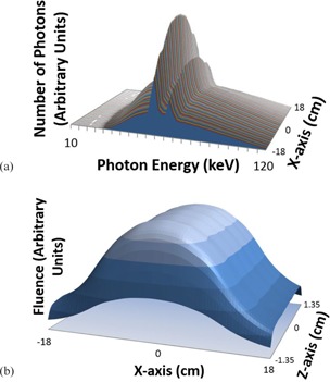
The purpose of this study was to show that the nominal peak tube voltage potential (kVp) and measured half-value layer (HVL) can be used to generate energy spectra and fluence profiles for characterizing a computed tomography (CT) X-ray source, and to validate the source model and an in-house kV X-ray dose computation algorithm (kVDoseCalc) for computing machine- and patient-specific CT dose. Spatial variation of the X-ray source spectra of a Philips Brilliance and a GE Optima Big Bore CT scanner were found by measuring the HVL along the direction of the internal bow-tie filter axes. Third-party software, Spektr, and the nominal kVp settings were used to generate the energy spectra. Beam fluence was calculated by dividing the integral product of the spectra and the in-air NIST mass-energy attenuation coefficients by in-air dose measurements along the filter axis. The authors found the optimal number of photons to seed in kVDoseCalc to achieve dose convergence. The Philips Brilliance beams were modeled for 90, 120, and 140 kVp tube settings. The GE Optima beams were modeled for 80, 100, 120, and 140 kVp tube settings. Relative doses measured using a Capintec Farmer-type ionization chamber (0.65 cc) placed in a cylindrical polymethyl methacrylate (PMMA) phantom and irradiated by the Philips Brilliance, were compared to those computed with kVDoseCalc. Relative doses in an anthropomorphic thorax phantom (E2E SBRT Phantom) irradiated by the GE Optima were measured using a (0.015 cc) PTW Freiburg ionization chamber and compared to computations from kVDoseCalc. The number of photons required to reduce the average statistical uncertainty in dose to < 0.3% was 2 × 10⁵. The average percent difference between calculation and measurement over all 12 PMMA phantom positions was found to be 1.44%, 1.47%, and 1.41% for 90, 120, and 140 kVp, respectively. The maximum percent difference between calculation and measurement for all energies, measurement positions, and phantoms was less than 3.50%. Thirty-five out of a total of 36 simulation conditions were within the experimental uncertainties associated with measurement reproducibility and chamber volume effects for the PMMA phantom. The agreement between calculation and measurement was within experimental uncertainty for 19 out of 20 simulation conditions at five points of interest in the anthropomorphic thorax phantom for the four beam energies modeled. The source model and characterization technique based on HVL measurements and nominal kVp can be used to accurately compute CT dose. This accuracy provides experimental validation of kVDoseCalc for computing CT dose.
本研究的目的是表明,标称峰值管电压(kVp)和测量的半值层(HVL)可用于生成能谱和注量分布,以表征计算机断层扫描(CT)X射线源,并验证用于计算特定机器和患者的CT剂量的源模型和内部kV X射线剂量计算算法(kVDoseCalc)。通过沿内部蝴蝶结滤波器轴方向测量HVL,发现了飞利浦Brilliance和GE Optima大孔径CT扫描仪的X射线源光谱的空间变化。使用第三方软件Spektr和标称kVp设置生成能谱。通过将光谱与空气中NIST质量能量衰减系数的积分乘积除以沿滤波器轴的空气中剂量测量值来计算束流注量。作者找到了在kVDoseCalc中播种以实现剂量收敛的最佳光子数。对飞利浦Brilliance光束在90、120和140 kVp管设置下进行了建模。对GE Optima光束在80、100、120和140 kVp管设置下进行了建模。将置于圆柱形聚甲基丙烯酸甲酯(PMMA)体模中并由飞利浦Brilliance照射的Capintec Farmer型电离室(0.65 cc)测量的相对剂量与用kVDoseCalc计算的剂量进行比较。使用(0.015 cc)PTW弗莱堡电离室测量GE Optima照射的人体胸部体模(E2E SBRT体模)中的相对剂量,并与kVDoseCalc的计算结果进行比较。将剂量平均统计不确定度降低到<0.3%所需的光子数为2×10⁵。在所有12个PMMA体模位置上,计算值与测量值之间的平均百分比差异在90、120和140 kVp时分别为1.44%、1.47%和1.41%。在所有能量、测量位置和体模下,计算值与测量值之间的最大百分比差异小于3.50%。在总共36个模拟条件中,有35个在与PMMA体模的测量再现性和电离室体积效应相关的实验不确定度范围内。在对四种束能量进行建模的人体胸部体模的五个感兴趣点处,20个模拟条件中有19个的计算值与测量值之间的一致性在实验不确定度范围内。基于HVL测量和标称kVp的源模型和表征技术可用于准确计算CT剂量。这种准确性为kVDoseCalc计算CT剂量提供了实验验证。