Schwede Angela, Macleod Olivia J S, MacGregor Paula, Carrington Mark
Department of Biochemistry, University of Cambridge, Cambridge, United Kingdom.
PLoS Pathog. 2015 Dec 31;11(12):e1005259. doi: 10.1371/journal.ppat.1005259. eCollection 2015 Dec.
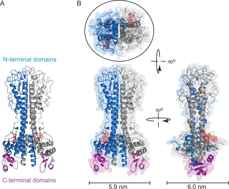
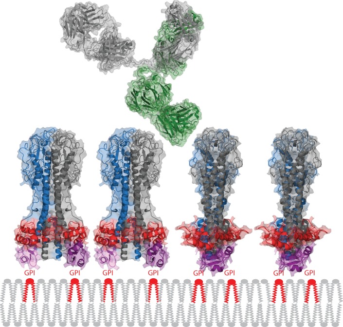
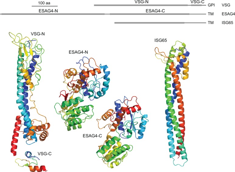
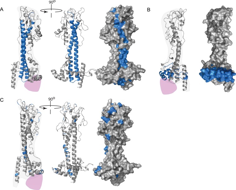
Variations on the statement "the variant surface glycoprotein (VSG) coat that covers the external face of the mammalian bloodstream form of Trypanosoma brucei acts a physical barrier" appear regularly in research articles and reviews. The concept of the impenetrable VSG coat is an attractive one, as it provides a clear model for understanding how a trypanosome population persists; each successive VSG protects the plasma membrane and is immunologically distinct from previous VSGs. What is the evidence that the VSG coat is an impenetrable barrier, and how do antibodies and other extracellular proteins interact with it? In this review, the nature of the extracellular surface of the bloodstream form trypanosome is described, and past experiments that investigated binding of antibodies and lectins to trypanosomes are analysed using knowledge of VSG sequence and structure that was unavailable when the experiments were performed. Epitopes for some VSG monoclonal antibodies are mapped as far as possible from previous experimental data, onto models of VSG structures. The binding of lectins to some, but not to other, VSGs is revisited with more recent knowledge of the location and nature of N-linked oligosaccharides. The conclusions are: (i) Much of the variation observed in earlier experiments can be explained by the identity of the individual VSGs. (ii) Much of an individual VSG is accessible to antibodies, and the barrier that prevents access to the cell surface is probably at the base of the VSG N-terminal domain, approximately 5 nm from the plasma membrane. This second conclusion highlights a gap in our understanding of how the VSG coat works, as several plasma membrane proteins with large extracellular domains are very unlikely to be hidden from host antibodies by VSG.
“覆盖布氏锥虫哺乳动物血流形式外表面的变异表面糖蛋白(VSG)衣壳起到物理屏障作用”这一表述的各种变体经常出现在研究文章和综述中。不可穿透的VSG衣壳这一概念很有吸引力,因为它为理解锥虫群体如何持续存在提供了一个清晰的模型;每一个相继的VSG都保护质膜,并且在免疫上与先前的VSG不同。VSG衣壳是不可穿透屏障的证据是什么,抗体和其他细胞外蛋白如何与之相互作用?在这篇综述中,描述了血流形式锥虫细胞外表面的性质,并利用实验进行时尚未获得的VSG序列和结构知识,分析了过去研究抗体和凝集素与锥虫结合的实验。根据先前的实验数据,尽可能将一些VSG单克隆抗体的表位映射到VSG结构模型上。结合N-连接寡糖的位置和性质的最新知识,重新审视了凝集素与某些但不是其他VSG的结合情况。结论如下:(i)早期实验中观察到的许多变异可以用单个VSG的特性来解释。(ii)单个VSG的大部分区域可被抗体识别,阻止抗体接近细胞表面的屏障可能位于VSG N末端结构域的基部,距质膜约5纳米。第二个结论凸显了我们在理解VSG衣壳如何发挥作用方面的差距,因为一些具有大细胞外结构域的质膜蛋白极不可能被VSG隐藏而不被宿主抗体识别。