Kalcher Klaudius, Boubela Roland N, Huf Wolfgang, Našel Christian, Moser Ewald
Center for Medical Physics and Biomedical Engineering, Medical University of ViennaVienna, Austria; MR Centre of Excellence, Medical University of ViennaVienna, Austria.
Department of Radiology, Tulln Hospital, Karl Landsteiner University of Health Sciences Tulln, Austria.
Front Neurosci. 2015 Dec 16;9:472. doi: 10.3389/fnins.2015.00472. eCollection 2015.
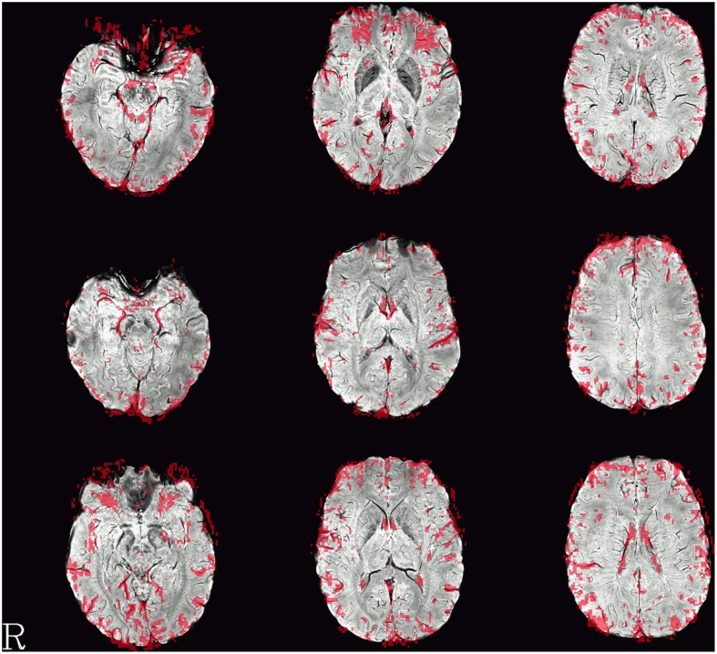
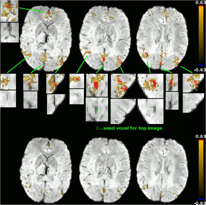
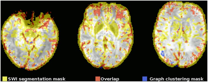
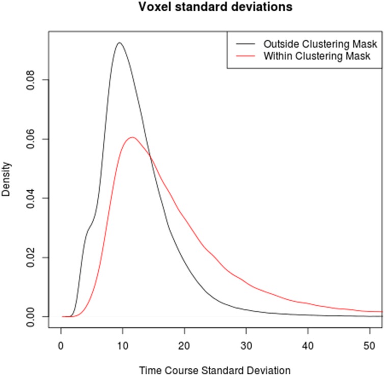
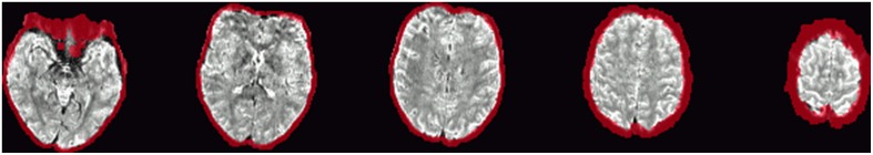
Identifying venous voxels in fMRI datasets is important to increase the specificity of fMRI analyses to microvasculature in the vicinity of the neural processes triggering the BOLD response. This is, however, difficult to achieve in particular in typical studies where magnitude images of BOLD EPI are the only data available. In this study, voxelwise functional connectivity graphs were computed on minimally preprocessed low TR (333 ms) multiband resting-state fMRI data, using both high positive and negative correlations to define edges between nodes (voxels). A high correlation threshold for binarization ensures that most edges in the resulting sparse graph reflect the high coherence of signals in medium to large veins. Graph clustering based on the optimization of modularity was then employed to identify clusters of coherent voxels in this graph, and all clusters of 50 or more voxels were then interpreted as corresponding to medium to large veins. Indeed, a comparison with SWI reveals that 75.6±5.9% of voxels within these large clusters overlap with veins visible in the SWI image or lie outside the brain parenchyma. Some of the remaining differences between the two modalities can be explained by imperfect alignment or geometric distortions between the two images. Overall, the graph clustering based method for identifying venous voxels has a high specificity as well as the additional advantages of being computed in the same voxel grid as the fMRI dataset itself and not needing any additional data beyond what is usually acquired (and exported) in standard fMRI experiments.
在功能磁共振成像(fMRI)数据集中识别静脉体素,对于提高fMRI分析对触发血氧水平依赖(BOLD)反应的神经过程附近微血管系统的特异性非常重要。然而,这在特别是典型研究中很难实现,在这些研究中,BOLD回波平面成像(EPI)的幅度图像是唯一可用的数据。在本研究中,基于最小预处理的低重复时间(TR,333毫秒)多波段静息态fMRI数据计算体素级功能连接图,使用高正相关和负相关来定义节点(体素)之间的边。用于二值化的高相关阈值确保了所得稀疏图中的大多数边反映了中到大静脉中信号的高相干性。然后采用基于模块度优化的图聚类来识别该图中相干体素的簇,然后将所有50个或更多体素的簇解释为对应于中到大静脉。事实上,与磁敏感加权成像(SWI)的比较表明,这些大簇内75.6±5.9%的体素与SWI图像中可见的静脉重叠或位于脑实质之外。两种模态之间其余的一些差异可以通过两幅图像之间的不完全对齐或几何畸变来解释。总体而言,基于图聚类的静脉体素识别方法具有很高的特异性,并且具有在与fMRI数据集本身相同的体素网格中计算以及除了标准fMRI实验中通常采集(和导出)的数据之外不需要任何额外数据的额外优点。