Marins Theo F, Rodrigues Erika C, Engel Annerose, Hoefle Sebastian, Basílio Rodrigo, Lent Roberto, Moll Jorge, Tovar-Moll Fernanda
D'Or Institute for Research and EducationRio de Janeiro, Brazil; Institute of Biomedical Sciences, Federal University of Rio de JaneiroRio de Janeiro, Brazil.
D'Or Institute for Research and EducationRio de Janeiro, Brazil; Augusto Motta University (Unisuam)Rio de Janeiro, Brazil.
Front Behav Neurosci. 2015 Dec 24;9:341. doi: 10.3389/fnbeh.2015.00341. eCollection 2015.
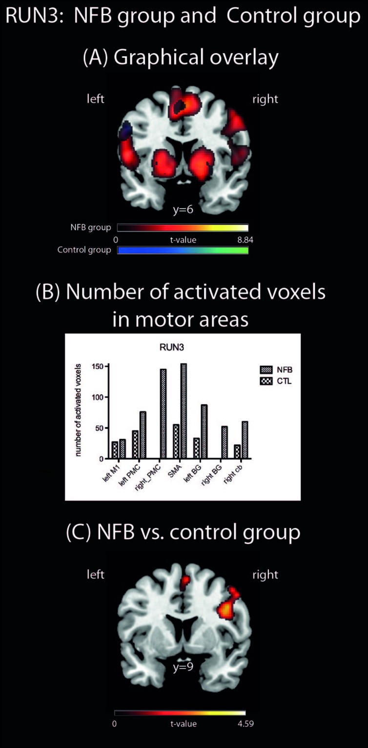
Neurofeedback by functional magnetic resonance imaging (fMRI) is a technique of potential therapeutic relevance that allows individuals to be aware of their own neurophysiological responses and to voluntarily modulate the activity of specific brain regions, such as the premotor cortex (PMC), important for motor recovery after brain injury. We investigated (i) whether healthy human volunteers are able to up-regulate the activity of the left PMC during a right hand finger tapping motor imagery (MI) task while receiving continuous fMRI-neurofeedback, and (ii) whether successful modulation of brain activity influenced non-targeted motor control regions. During the MI task, participants of the neurofeedback group (NFB) received ongoing visual feedback representing the level of fMRI responses within their left PMC. Control (CTL) group participants were shown similar visual stimuli, but these were non-contingent on brain activity. Both groups showed equivalent levels of behavioral ratings on arousal and MI, before and during the fMRI protocol. In the NFB, but not in CLT group, brain activation during the last run compared to the first run revealed increased activation in the left PMC. In addition, the NFB group showed increased activation in motor control regions extending beyond the left PMC target area, including the supplementary motor area, basal ganglia and cerebellum. Moreover, in the last run, the NFB group showed stronger activation in the left PMC/inferior frontal gyrus when compared to the CTL group. Our results indicate that modulation of PMC and associated motor control areas can be achieved during a single neurofeedback-fMRI session. These results contribute to a better understanding of the underlying mechanisms of MI-based neurofeedback training, with direct implications for rehabilitation strategies in severe brain disorders, such as stroke.
基于功能磁共振成像(fMRI)的神经反馈是一种具有潜在治疗意义的技术,它使个体能够意识到自己的神经生理反应,并自愿调节特定脑区的活动,如对脑损伤后运动恢复很重要的运动前皮质(PMC)。我们研究了:(i)健康人类志愿者在接受连续fMRI神经反馈时,是否能够在右手手指敲击运动想象(MI)任务期间上调左侧PMC的活动;(ii)脑活动的成功调节是否会影响非目标运动控制区域。在MI任务期间,神经反馈组(NFB)的参与者收到了代表其左侧PMC内fMRI反应水平的实时视觉反馈。对照组(CTL)的参与者看到了类似的视觉刺激,但这些刺激与脑活动无关。在fMRI检查之前和期间,两组在唤醒和MI方面的行为评分水平相当。在NFB组而非CLT组中,与第一次扫描相比,最后一次扫描期间的脑激活显示左侧PMC的激活增加。此外,NFB组在超出左侧PMC目标区域的运动控制区域,包括辅助运动区、基底神经节和小脑,也显示出激活增加。此外,在最后一次扫描中,与CTL组相比,NFB组在左侧PMC/额下回显示出更强的激活。我们的结果表明,在单次神经反馈fMRI训练期间,可以实现对PMC和相关运动控制区域的调节。这些结果有助于更好地理解基于MI的神经反馈训练的潜在机制,对中风等严重脑部疾病的康复策略有直接影响。