Department of Pediatrics Vanderbilt University Nashville Tennessee.
Department of Biomedical Engineering Vanderbilt University Nashville Tennessee.
Ann Clin Transl Neurol. 2015 Oct 27;2(12):1041-54. doi: 10.1002/acn3.254. eCollection 2015 Dec.
While abnormalities in myelin in tuberous sclerosis complex (TSC) have been known for some time, recent imaging-based data suggest myelin abnormalities may be independent of the pathognomonic cortical lesions ("tubers"). Multiple mouse models of TSC exhibit myelination deficits, though the cell types responsible and the mechanisms underlying the myelin abnormalities remain unclear.
To determine the role of alterations in mTOR signaling in myelination, we generated a conditional knockout (CKO) mouse model using Cre-recombinase and the Olig2 promoter to inactivate the Tsc2 gene in oligodendrocyte precursor cells.
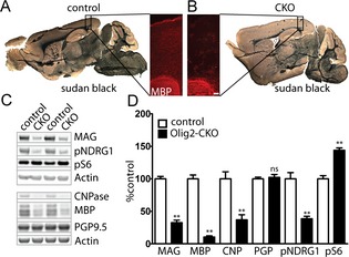
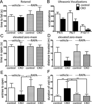
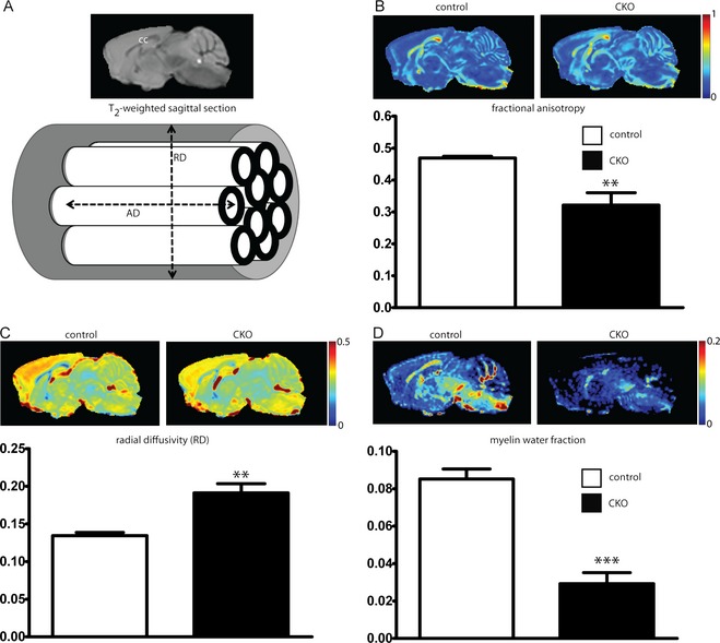
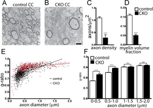
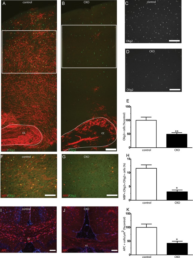
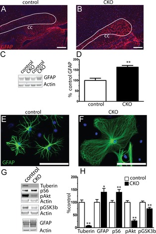
Characterization of myelin and myelin constituent proteins demonstrated a marked hypomyelination phenotype. Diffusion-based magnetic resonance imaging studies were likewise consistent with hypomyelination. Hypomyelination was due in part to decreased myelinated axon density and myelin thickness as well as decreased oligodendrocyte numbers. Coincident with hypomyelination, an extensive gliosis was seen in both the cortex and white matter tracks, suggesting alterations in cell fate due to changes in mTOR activity in oligodendrocyte precursors. Despite a high-frequency appendicular tremor and altered gait in CKO mice, no significant changes in activity, vocalizations, or anxiety-like phenotypes were seen.
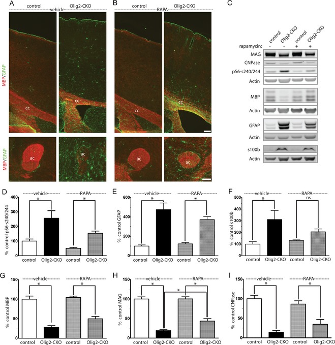
Our findings support a known role of mTOR signaling in regulation of myelination and demonstrate that increased mTORC1 activity early in development within oligodendrocytes results in hypomyelination and not hypermyelination. Our data further support a dissociation between decreased Akt activity and increased mTORC1 activity toward hypomyelination. Thus, therapies promoting activation of Akt-dependent pathways while reducing mTORC1 activity may prove beneficial in treatment of human disease.
虽然结节性硬化症(TSC)中的髓鞘异常已经为人所知一段时间,但最近基于成像的研究数据表明,髓鞘异常可能与特有的皮质病变(“结节”)无关。多种 TSC 小鼠模型均表现出髓鞘缺陷,但导致髓鞘异常的细胞类型以及潜在机制仍不清楚。
为了确定 mTOR 信号转导改变在髓鞘形成中的作用,我们使用 Cre 重组酶和 Olig2 启动子生成了条件敲除(CKO)小鼠模型,以在少突胶质前体细胞中使 Tsc2 基因失活。
髓鞘和髓鞘组成蛋白的特征表明存在明显的少突胶质细胞发育不全表型。基于扩散的磁共振成像研究也与少突胶质细胞发育不全一致。少突胶质细胞发育不全部分归因于少突胶质细胞前体细胞中 mTOR 活性改变导致有髓神经轴突密度和髓鞘厚度降低以及少突胶质细胞数量减少。与少突胶质细胞发育不全相一致,在皮质和白质轨迹中均观察到广泛的神经胶质增生,这表明由于 mTOR 活性改变,少突胶质细胞前体细胞的细胞命运发生改变。尽管 CKO 小鼠出现高频肢体震颤和步态改变,但在活动、发声或焦虑样表型方面未见明显变化。
我们的研究结果支持 mTOR 信号在调节髓鞘形成中的已知作用,并表明在发育早期增加少突胶质细胞中的 mTORC1 活性会导致少突胶质细胞发育不全,而不是髓鞘过度形成。我们的数据进一步支持 Akt 活性降低和 mTORC1 活性增加与少突胶质细胞发育不全之间的分离。因此,促进 Akt 依赖性途径激活同时降低 mTORC1 活性的疗法可能对人类疾病的治疗有益。