Ontsouka Edgar Corneille, Bertschi Janique Sabina, Huang Xiao, Lüthi Michael, Müller Stefan, Albrecht Christiane
Faculty of Medicine, Institute of Biochemistry and Molecular Medicine, University of Bern, Buehlstrasse 28, 3012, Bern, Switzerland.
Swiss National Center of Competence in Research, NCCR TransCure, University of Bern, Bern, Switzerland.
Biol Res. 2016 Jan 6;49:1. doi: 10.1186/s40659-015-0063-2.
Mammary cell cultures are convenient tools for in vitro studies of mammary gland biology. However, the heterogeneity of mammary cell types, e.g., glandular milk secretory epithelial or myoepithelial cells, often complicates the interpretation of cell-based data. The present study was undertaken to determine the relevance of bovine primary mammary epithelial cells isolated from American Holstein (bMECUS) or Swiss Holstein-Friesian (bMECCH) cows, and of primary bovine mammary alveolar epithelial cells stably transfected with simian virus-40 (SV-40) large T-antigen (MAC-T) for in vitro analyses. This was evaluated by testing their expression pattern of cytokeratin (CK) 7, 18, 19, vimentin, and α-smooth muscle actin (α-SMA).
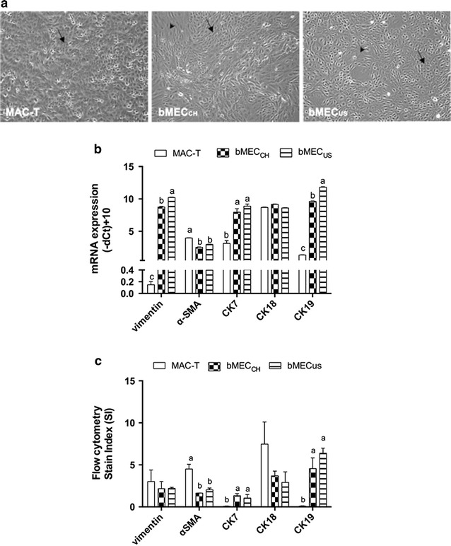
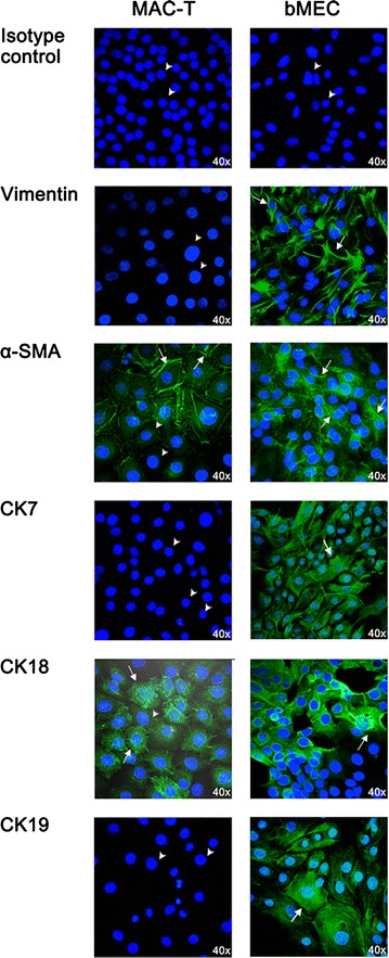
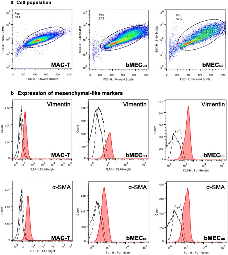
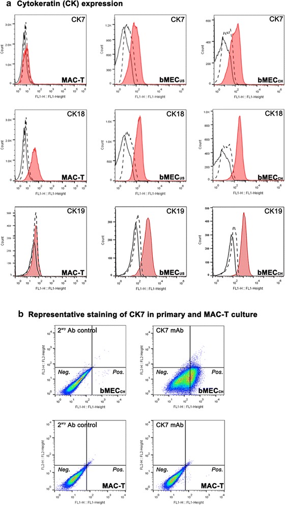
The expression of the listed markers was assessed using real-time quantitative PCR, flow cytometry and immunofluorescence microscopy. Characteristic markers of the mesenchymal (vimentin), myoepithelial (α-SMA) and glandular secretory cells (CKs) showed differential expression among the studied cell cultures, partly depending on the analytical method used. The relative mRNA expression of vimentin, CK7 and CK19, respectively, was lower (P < 0.05) in immortalized than in primary mammary cell cultures. The stain index (based on flow cytometry) of CK7 and CK19 protein was lower (P < 0.05) in MAC-T than in bMECs, while the expression of α-SMA and CK18 showed an inverse pattern. Immunofluorescence microscopy analysis mostly confirmed the mRNA data, while partly disagreed with flow cytometry data (e.g., vimentin level in MAC-T). The differential expression of CK7 and CK19 allowed discriminating between immortal and primary mammary cultures.
The expression of the selected widely used cell type markers in primary and immortalized MEC cells did not allow a clear preference between these two cell models for in vitro analyses studying aspects of milk composition. All tested cell models exhibited to a variable degree epithelial and mesenchymal features. Thus, based on their characterization with widely used cell markers, none of these cultures represent an unequivocal alveolar mammary epithelial cell model. For choosing the appropriate in vitro model additional properties such as the expression profile of specific proteins of interest (e.g., transporter proteins) should equally be taken into account.
乳腺细胞培养是乳腺生物学体外研究的便捷工具。然而,乳腺细胞类型的异质性,例如腺泡分泌上皮细胞或肌上皮细胞,常常使基于细胞的数据解释变得复杂。本研究旨在确定从美国荷斯坦奶牛(bMECUS)或瑞士荷斯坦 - 弗里生奶牛(bMECCH)分离的牛原代乳腺上皮细胞,以及稳定转染猿猴病毒40(SV - 40)大T抗原的原代牛乳腺腺泡上皮细胞(MAC - T)用于体外分析的相关性。通过检测它们细胞角蛋白(CK)7、18、19、波形蛋白和α - 平滑肌肌动蛋白(α - SMA)的表达模式来进行评估。
使用实时定量PCR、流式细胞术和免疫荧光显微镜评估所列标志物的表达。间充质细胞(波形蛋白)、肌上皮细胞(α - SMA)和腺泡分泌细胞(CKs)的特征性标志物在研究的细胞培养物中表现出差异表达,部分取决于所使用的分析方法。波形蛋白、CK7和CK19的相对mRNA表达在永生化乳腺细胞培养物中分别低于原代乳腺细胞培养物(P < 0.05)。MAC - T中CK7和CK19蛋白的染色指数(基于流式细胞术)低于bMECs(P < 0.05),而α - SMA和CK18的表达呈现相反模式。免疫荧光显微镜分析大多证实了mRNA数据,但部分与流式细胞术数据不一致(例如MAC - T中的波形蛋白水平)。CK7和CK19的差异表达使得能够区分永生化和原代乳腺培养物。
在原代和永生化乳腺上皮细胞中,所选广泛使用的细胞类型标志物的表达并不能明确表明在研究乳汁成分方面的体外分析中这两种细胞模型哪一种更具优势。所有测试的细胞模型都不同程度地表现出上皮和间充质特征。因此,基于它们用广泛使用的细胞标志物进行的表征,这些培养物中没有一种代表明确的腺泡乳腺上皮细胞模型。为了选择合适的体外模型,还应同样考虑其他特性,例如感兴趣的特定蛋白质(例如转运蛋白)的表达谱。