Wang Ellen F, Misra Stuti L, Patel Dipika V
Department of Ophthalmology, New Zealand National Eye Centre, Faculty of Medical and Health Sciences, The University of Auckland, Auckland 1142, New Zealand.
Biomed Res Int. 2015;2015:951081. doi: 10.1155/2015/951081. Epub 2015 Dec 7.
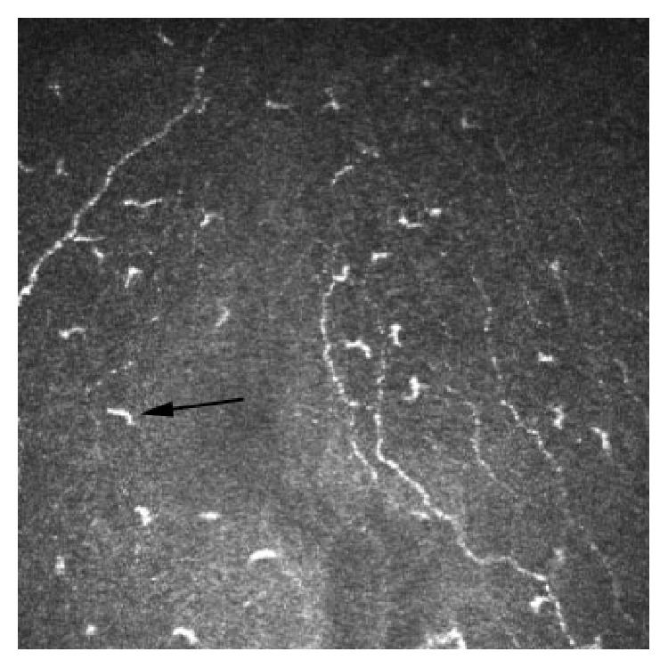
In vivo confocal microscopy (IVCM) of the living human cornea offers the ability to perform repeated imaging without tissue damage. Studies using corneal IVCM have led to significant contributions to scientific and clinical knowledge of the living cornea in health and pathological states. Recently the application of corneal IVCM beyond ophthalmology to wider clinical and research fields has been demonstrated. Abnormalities of the corneal subbasal nerve plexus have been associated with many forms of peripheral neuropathy and Langerhans cells correlate with systemic inflammatory states. There is a rapidly growing evidence base investigating the use of corneal IVCM in many systemic conditions and a well-established evidence base for IVCM imaging of the corneal subbasal plexus in diabetic peripheral neuropathy. This paper reviews the potential use of corneal IVCM in general clinical practice as a noninvasive method of assessing peripheral neuropathies, monitoring inflammatory states and clinical therapeutic response.
对活人角膜进行的体内共聚焦显微镜检查(IVCM)能够在不造成组织损伤的情况下进行重复成像。使用角膜IVCM的研究为健康和病理状态下活人角膜的科学及临床知识做出了重大贡献。最近,已证明角膜IVCM在眼科以外的更广泛临床和研究领域中的应用。角膜基底神经丛异常与多种形式的周围神经病变相关,而朗格汉斯细胞与全身炎症状态相关。越来越多的证据表明角膜IVCM可用于多种全身性疾病,并且在糖尿病性周围神经病变中角膜基底丛IVCM成像有成熟的证据基础。本文综述了角膜IVCM在一般临床实践中作为评估周围神经病变、监测炎症状态和临床治疗反应的非侵入性方法的潜在用途。