• 文献检索
  • 文档翻译
  • 深度研究
  • 学术资讯
  • Suppr Zotero 插件Zotero 插件
  • 邀请有礼
  • 套餐&价格
  • 历史记录
应用&插件
Suppr Zotero 插件Zotero 插件浏览器插件Mac 客户端Windows 客户端微信小程序
定价
高级版会员购买积分包购买API积分包
服务
文献检索文档翻译深度研究API 文档MCP 服务
关于我们
关于 Suppr公司介绍联系我们用户协议隐私条款
关注我们

Suppr 超能文献

核心技术专利:CN118964589B侵权必究
粤ICP备2023148730 号-1Suppr @ 2026

文献检索

告别复杂PubMed语法,用中文像聊天一样搜索,搜遍4000万医学文献。AI智能推荐,让科研检索更轻松。

立即免费搜索

文件翻译

保留排版,准确专业,支持PDF/Word/PPT等文件格式,支持 12+语言互译。

免费翻译文档

深度研究

AI帮你快速写综述,25分钟生成高质量综述,智能提取关键信息,辅助科研写作。

立即免费体验

新型广角活体角膜共焦显微镜与皮肤活检评估 2 型糖尿病周围神经病变的比较。

Comparison of Novel Wide-Field In Vivo Corneal Confocal Microscopy With Skin Biopsy for Assessing Peripheral Neuropathy in Type 2 Diabetes.

机构信息

Department of Medical Biochemistry, Unit of Regenerative Medicine, Oslo University Hospital, Oslo, Norway.

Department of Translational Medicine, Hand Surgery, Lund University, Malmö, Sweden.

出版信息

Diabetes. 2023 Jul 1;72(7):908-917. doi: 10.2337/db22-0863.

DOI:10.2337/db22-0863
PMID:37058418
原文链接:https://pmc.ncbi.nlm.nih.gov/articles/PMC10281223/
Abstract

UNLABELLED

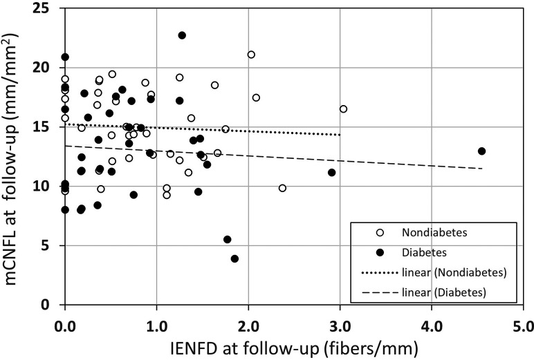
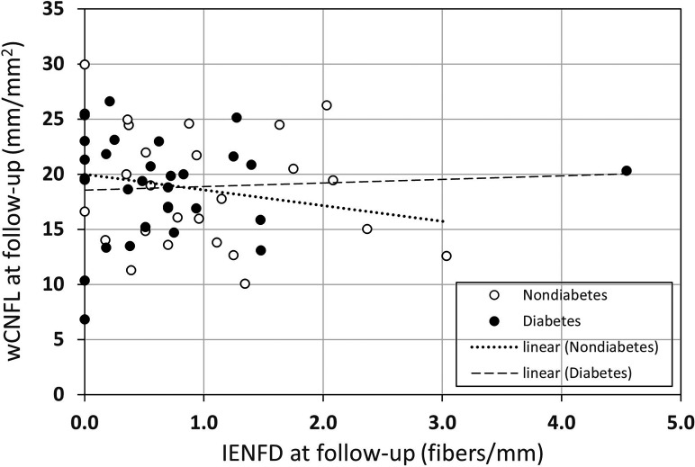
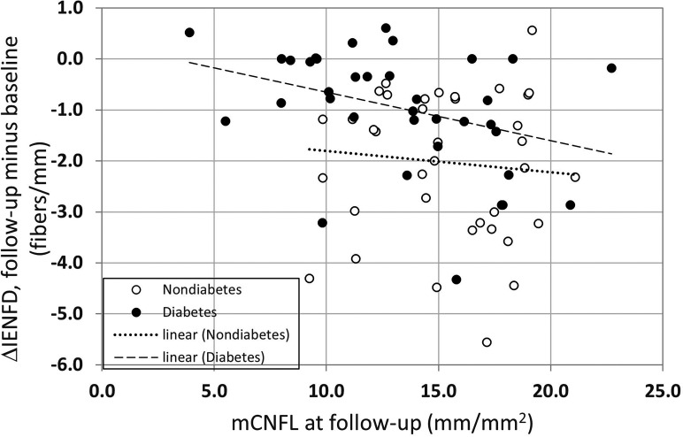
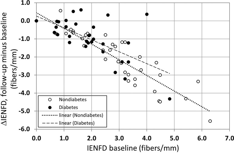
Diabetic peripheral neuropathy (DPN) is a serious complication of diabetes, where skin biopsy assessing intraepidermal nerve fiber density (IENFD) plays an important diagnostic role. In vivo confocal microscopy (IVCM) of the corneal subbasal nerve plexus has been proposed as a noninvasive diagnostic modality for DPN. Direct comparisons of skin biopsy and IVCM in controlled cohorts are lacking, as IVCM relies on subjective selection of images depicting only 0.2% of the nerve plexus. We compared these diagnostic modalities in a fixed-age cohort of 41 participants with type 2 diabetes and 36 healthy participants using machine algorithms to create wide-field image mosaics and quantify nerves in an area 37 times the size of prior studies to avoid human bias. In the same participants, and at the same time point, no correlation between IENFD and corneal nerve density was found. Corneal nerve density did not correlate with clinical measures of DPN, including neuropathy symptom and disability scores, nerve conduction studies, or quantitative sensory tests. Our findings indicate that corneal and intraepidermal nerves likely mirror different aspects of nerve degeneration, where only intraepidermal nerves appear to reflect the clinical status of DPN, suggesting that scrutiny is warranted concerning methodologies of studies using corneal nerves to assess DPN.

ARTICLE HIGHLIGHTS

Comparison of intraepidermal nerve fiber density with automated wide-field corneal nerve fiber density in participants with type 2 diabetes revealed no correlation between these parameters. Intraepidermal and corneal nerve fibers both detected neurodegeneration in type 2 diabetes, but only intraepidermal nerve fibers were associated with clinical measures of diabetic peripheral neuropathy. A lack of association of corneal nerves with peripheral neuropathy measures suggests that corneal nerve fibers may be a poor biomarker for diabetic peripheral neuropathy.

摘要

目的

描述糖尿病患者角膜神经纤维密度与皮肤神经纤维密度的相关性。

方法

在这项横断面研究中,我们纳入了 41 名 2 型糖尿病患者和 36 名健康对照者,采集他们的皮肤和角膜活检标本,以评估皮肤内神经纤维密度(IENFD)和角膜神经纤维密度(CNFD)。我们使用机器算法创建了宽视野图像镶嵌图,并对之前研究面积的 37 倍的区域进行神经定量,以避免人为偏差。

结果

在所有参与者中,IENFD 与 CNFD 之间均无相关性。CNFD 与 DPN 的临床测量值(包括神经病变症状和残疾评分、神经传导研究或定量感觉测试)均无相关性。

结论

这些发现表明,角膜和表皮内神经可能反映了神经退化的不同方面,只有表皮内神经似乎反映了 DPN 的临床状况,这表明使用角膜神经评估 DPN 的研究方法需要仔细审查。

https://cdn.ncbi.nlm.nih.gov/pmc/blobs/87f2/10281223/985b8c83c126/db220863f5.jpg
https://cdn.ncbi.nlm.nih.gov/pmc/blobs/87f2/10281223/6be3c1053852/db220863f1.jpg
https://cdn.ncbi.nlm.nih.gov/pmc/blobs/87f2/10281223/9671572bfc92/db220863f2.jpg
https://cdn.ncbi.nlm.nih.gov/pmc/blobs/87f2/10281223/e12263ed345f/db220863f3.jpg
https://cdn.ncbi.nlm.nih.gov/pmc/blobs/87f2/10281223/56f2cd4fb5f7/db220863f4.jpg
https://cdn.ncbi.nlm.nih.gov/pmc/blobs/87f2/10281223/985b8c83c126/db220863f5.jpg

相似文献

1
Comparison of Novel Wide-Field In Vivo Corneal Confocal Microscopy With Skin Biopsy for Assessing Peripheral Neuropathy in Type 2 Diabetes.新型广角活体角膜共焦显微镜与皮肤活检评估 2 型糖尿病周围神经病变的比较。
Diabetes. 2023 Jul 1;72(7):908-917. doi: 10.2337/db22-0863.
2
Assessing Corneal Confocal Microscopy and Other Small Fiber Measures in Diabetic Polyneuropathy.评估糖尿病性多发性神经病中的角膜共焦显微镜和其他小纤维测量方法。
Neurology. 2023 Apr 18;100(16):e1680-e1690. doi: 10.1212/WNL.0000000000206902. Epub 2023 Feb 7.
3
Utility of corneal confocal microscopy for assessing mild diabetic neuropathy: baseline findings of the LANDMark study.角膜共焦显微镜在评估轻度糖尿病神经病变中的应用:LANDMark研究的基线结果
Clin Exp Optom. 2012 May;95(3):348-54. doi: 10.1111/j.1444-0938.2012.00740.x. Epub 2012 Apr 29.
4
Retinal and Corneal Neurodegeneration and Their Association with Systemic Signs of Peripheral Neuropathy in Type 2 Diabetes.视网膜和角膜神经退行性变及其与 2 型糖尿病周围神经病变系统表现的关系。
Am J Ophthalmol. 2020 Jan;209:197-205. doi: 10.1016/j.ajo.2019.09.010. Epub 2019 Sep 19.
5
Imaging of the Corneal Subbasal Whorl-like Nerve Plexus: More Accurate Depiction of the Extent of Corneal Nerve Damage in Patients With Diabetes.角膜基底膜下涡状神经丛成像:更准确描绘糖尿病患者角膜神经损伤程度
Invest Ophthalmol Vis Sci. 2015 Aug;56(9):5417-23. doi: 10.1167/iovs.15-16609.
6
A Comparative Study on the Diagnostic Utility of Corneal Confocal Microscopy and Tear Neuromediator Levels in Diabetic Peripheral Neuropathy.角膜共焦显微镜与泪神经递质水平在糖尿病周围神经病变诊断中的对比研究。
Curr Eye Res. 2020 Aug;45(8):921-930. doi: 10.1080/02713683.2019.1705984. Epub 2019 Dec 26.
7
Early detection of nerve fiber loss by corneal confocal microscopy and skin biopsy in recently diagnosed type 2 diabetes.角膜共焦显微镜和皮肤活检对近期诊断的 2 型糖尿病患者神经纤维丢失的早期检测。
Diabetes. 2014 Jul;63(7):2454-63. doi: 10.2337/db13-1819. Epub 2014 Feb 26.
8
Diagnostic utility of corneal confocal microscopy in type 2 diabetic peripheral neuropathy.角膜共焦显微镜在 2 型糖尿病周围神经病变中的诊断价值。
J Diabetes Investig. 2021 Apr;12(4):574-582. doi: 10.1111/jdi.13381. Epub 2020 Sep 25.
9
Corneal nerves in diabetes-The role of the in vivo corneal confocal microscopy of the subbasal nerve plexus in the assessment of peripheral small fiber neuropathy.糖尿病患者的角膜神经——眼表活体共聚焦显微镜在评估周围小纤维神经病变中的亚基底神经丛的作用。
Surv Ophthalmol. 2021 May-Jun;66(3):493-513. doi: 10.1016/j.survophthal.2020.09.003. Epub 2020 Sep 19.
10
Early nerve fibre regeneration in individuals with type 1 diabetes after simultaneous pancreas and kidney transplantation.1 型糖尿病患者胰肾联合移植后早期神经纤维再生。
Diabetologia. 2019 Aug;62(8):1478-1487. doi: 10.1007/s00125-019-4897-y. Epub 2019 Jun 7.

引用本文的文献

1
Methods for the evaluation of corneal nerve fibres in diabetes mellitus by in vivo confocal microscopy: a scoping review.通过体内共聚焦显微镜评估糖尿病患者角膜神经纤维的方法:一项范围综述
BMJ Open. 2025 Sep 4;15(9):e098294. doi: 10.1136/bmjopen-2024-098294.
2
Corneal Nerve Morphology in Painful Diabetic Neuropathy: A Meta-Analysis of In Vivo Confocal Microscopy Studies.疼痛性糖尿病神经病变中的角膜神经形态学:一项共聚焦显微镜活体研究的荟萃分析
Biomedicines. 2025 Jul 8;13(7):1675. doi: 10.3390/biomedicines13071675.
3
ConNeCT: weakly supervised corneal confocal microscopy image inpainting network based on a diffusion model.

本文引用的文献

1
Mosaic vs. Single Image Analysis with Confocal Microscopy of the Corneal Nerve Plexus for Diagnosis of Early Diabetic Peripheral Neuropathy.用于早期糖尿病周围神经病变诊断的角膜神经丛共聚焦显微镜检查的镶嵌图像与单图像分析对比
Ophthalmol Ther. 2022 Dec;11(6):2211-2223. doi: 10.1007/s40123-022-00574-z. Epub 2022 Oct 3.
2
Corneal and Epidermal Nerve Quantification in Chemotherapy Induced Peripheral Neuropathy.化疗所致周围神经病变中角膜和表皮神经的定量分析
Front Med (Lausanne). 2022 Feb 18;9:832344. doi: 10.3389/fmed.2022.832344. eCollection 2022.
3
Wide-field mosaics of the corneal subbasal nerve plexus in Parkinson's disease using in vivo confocal microscopy.
ConNeCT:基于扩散模型的弱监督角膜共焦显微镜图像修复网络
Biomed Opt Express. 2025 Jun 9;16(7):2615-2630. doi: 10.1364/BOE.562924. eCollection 2025 Jul 1.
4
Peripheral Neuropathy Symptoms and Ocular Surface Lesions in Patients with Type 2 Diabetes Mellitus and Dry Eye: A Clinical Correlational Study.2型糖尿病合并干眼症患者的周围神经病变症状与眼表病变:一项临床相关性研究
Ophthalmol Ther. 2025 May 15. doi: 10.1007/s40123-025-01150-x.
5
Automated program using convolutional neural networks for objective and reproducible selection of corneal confocal microscopy images.使用卷积神经网络的自动化程序用于角膜共焦显微镜图像的客观且可重复选择。
Digit Health. 2025 Mar 17;11:20552076251326223. doi: 10.1177/20552076251326223. eCollection 2025 Jan-Dec.
6
Research progress of corneal characteristics and changes in primary angle‑closure glaucoma (Review).原发性闭角型青光眼角膜特征及变化的研究进展(综述)
Biomed Rep. 2025 Feb 5;22(4):59. doi: 10.3892/br.2025.1937. eCollection 2025 Apr.
7
Quantifying the Corneal Nerve Whorl Pattern.量化角膜神经螺旋模式。
Transl Vis Sci Technol. 2024 Dec 2;13(12):11. doi: 10.1167/tvst.13.12.11.
8
Diabetic Peripheral Neuropathy: Emerging Treatments of Neuropathic Pain and Novel Diagnostic Methods.糖尿病性周围神经病变:神经性疼痛的新兴治疗方法及新型诊断方法
J Diabetes Sci Technol. 2024 Sep 16:19322968241279553. doi: 10.1177/19322968241279553.
9
Advances in diagnostic methods for early-stage diabetic polyneuropathy.早期糖尿病多发性神经病的诊断方法进展。
J Diabetes Investig. 2024 Jul;15(7):820-822. doi: 10.1111/jdi.14201. Epub 2024 Apr 6.
10
Applications of Imaging Technologies in Fuchs Endothelial Corneal Dystrophy: A Narrative Literature Review.成像技术在富克斯内皮性角膜营养不良中的应用:一篇叙述性文献综述
Bioengineering (Basel). 2024 Mar 11;11(3):271. doi: 10.3390/bioengineering11030271.
应用活体共聚焦显微镜观察帕金森病患者角膜基底部神经丛的广角镶嵌图像。
Sci Data. 2021 Nov 26;8(1):306. doi: 10.1038/s41597-021-01087-3.
4
Screening, diagnosis and management of diabetic sensorimotor polyneuropathy in clinical practice: International expert consensus recommendations.临床实践中糖尿病性感觉运动性多发性神经病的筛查、诊断与管理:国际专家共识推荐
Diabetes Res Clin Pract. 2022 Apr;186:109063. doi: 10.1016/j.diabres.2021.109063. Epub 2021 Sep 20.
5
Temporal trend of small nerve fibre degeneration in people with and without type 2 diabetes mellitus.2 型糖尿病患者与非糖尿病患者小神经纤维变性的时间趋势。
Diabet Med. 2022 Mar;39(3):e14691. doi: 10.1111/dme.14691. Epub 2021 Sep 22.
6
Painful diabetic neuropathy is associated with increased nerve regeneration in patients with type 2 diabetes undergoing intensive glycemic control.疼痛性糖尿病周围神经病变与 2 型糖尿病患者强化血糖控制后神经再生增加有关。
J Diabetes Investig. 2021 Sep;12(9):1642-1650. doi: 10.1111/jdi.13544. Epub 2021 May 3.
7
Corneal confocal microscopy identifies small fibre damage and progression of diabetic neuropathy.角膜共焦显微镜可识别小纤维损伤和糖尿病神经病变的进展。
Sci Rep. 2021 Jan 21;11(1):1859. doi: 10.1038/s41598-021-81302-8.
8
Corneal confocal microscopy compared with quantitative sensory testing and nerve conduction for diagnosing and stratifying the severity of diabetic peripheral neuropathy.角膜共焦显微镜检查与定量感觉测试及神经传导检查在诊断和分层糖尿病性周围神经病变严重程度方面的比较
BMJ Open Diabetes Res Care. 2020 Dec;8(2). doi: 10.1136/bmjdrc-2020-001801.
9
A Systematic Review of the Diagnostic Methods of Small Fiber Neuropathies in Rehabilitation.康复中小纤维神经病变诊断方法的系统评价
Diagnostics (Basel). 2020 Aug 20;10(9):613. doi: 10.3390/diagnostics10090613.
10
Prevalence and risk factors for diabetic neuropathy and painful diabetic neuropathy in primary and secondary healthcare in Qatar.卡塔尔初级和二级保健中糖尿病周围神经病变和痛性糖尿病周围神经病变的患病率和危险因素。
J Diabetes Investig. 2021 Apr;12(4):592-600. doi: 10.1111/jdi.13388. Epub 2020 Sep 14.