Moy Matthew P, Sauk Jenny, Gee Michael S
Division of Abdominal Imaging, Massachusetts General Hospital, Harvard Medical School, Boston, MA, USA; Division of Pediatric Imaging, Massachusetts General Hospital, Harvard Medical School, Boston, MA, USA.
Division of Gastroenterology, Massachusetts General Hospital, Harvard Medical School, Boston, MA, USA.
Gastroenterol Res Pract. 2016;2016:8168695. doi: 10.1155/2016/8168695. Epub 2015 Dec 27.
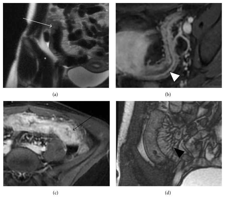
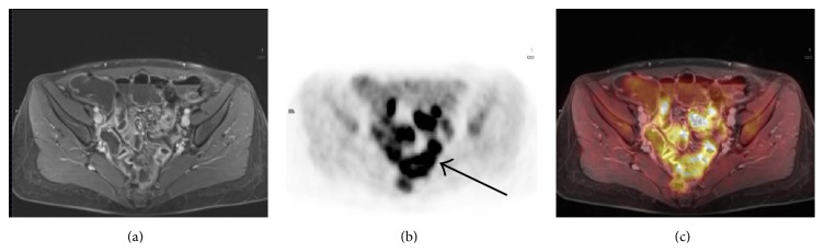
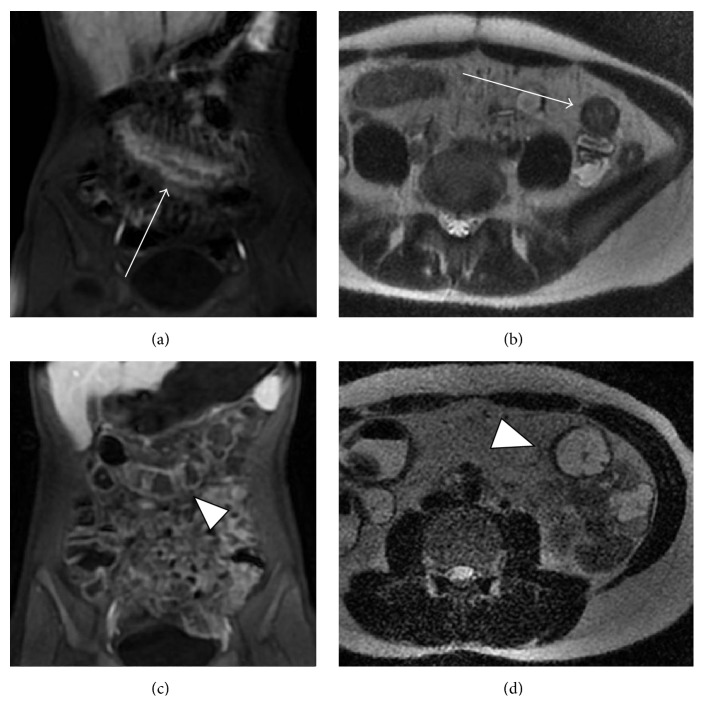
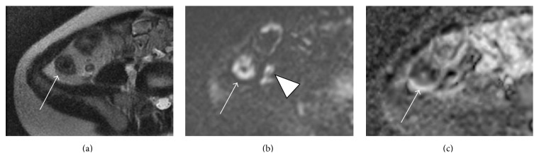
MR enterography (MRE) has become the primary imaging modality in the assessment of Crohn's disease (CD) in both children and adults at many institutions in the United States and worldwide, primarily due to its noninvasiveness, superior soft tissue contrast, and lack of ionizing radiation. MRE technique includes distention of the small bowel with oral contrast media with the acquisition of T2-weighted, balanced steady-state free precession, and multiphase T1-weighted fat suppressed gadolinium contrast-enhanced sequences. With the introduction of molecule-targeted biologic agents into the clinical setting for CD and their potential to reverse the inflammatory process, MRE is increasingly utilized to evaluate disease activity and response to therapy as an imaging complement to clinical indices or optical endoscopy. New and emerging MRE techniques, such as diffusion-weighted imaging (DWI), magnetization transfer, ultrasmall superparamagnetic iron oxide- (USPIO-) enhanced MRI, and PET-MR, offer the potential for an expanded role of MRI in detecting occult disease activity, evaluating early treatment response/resistance, and differentiating inflammatory from fibrotic strictures. Familiarity with MR enterography is essential for radiologists and gastroenterologists as the technique evolves and is further incorporated into the clinical management of CD.
在美国和全球的许多机构中,磁共振小肠造影(MRE)已成为评估儿童和成人克罗恩病(CD)的主要成像方式,这主要归功于其非侵入性、出色的软组织对比度以及无电离辐射。MRE技术包括通过口服对比剂使小肠扩张,并采集T2加权、平衡稳态自由进动和多期T1加权脂肪抑制钆对比增强序列。随着分子靶向生物制剂引入CD的临床治疗以及它们逆转炎症过程的潜力,MRE越来越多地被用作临床指标或光学内镜检查的成像补充手段,用于评估疾病活动度和对治疗的反应。新出现的MRE技术,如扩散加权成像(DWI)、磁化传递、超小超顺磁性氧化铁(USPIO)增强磁共振成像和PET-MR,为磁共振成像在检测隐匿性疾病活动、评估早期治疗反应/耐药性以及区分炎症性狭窄和纤维化狭窄方面发挥更大作用提供了可能。随着该技术的不断发展并进一步纳入CD的临床管理,放射科医生和胃肠病学家熟悉磁共振小肠造影至关重要。