Department of Radiology, Guy's & St Thomas' Hospital, Westminster Bridge Road, London, SE1 7EH, UK,
Insights Imaging. 2012 Jun;3(3):251-63. doi: 10.1007/s13244-012-0154-3. Epub 2012 Mar 17.
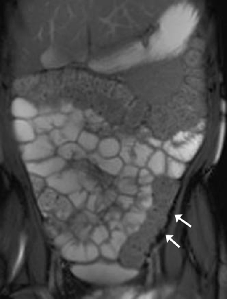
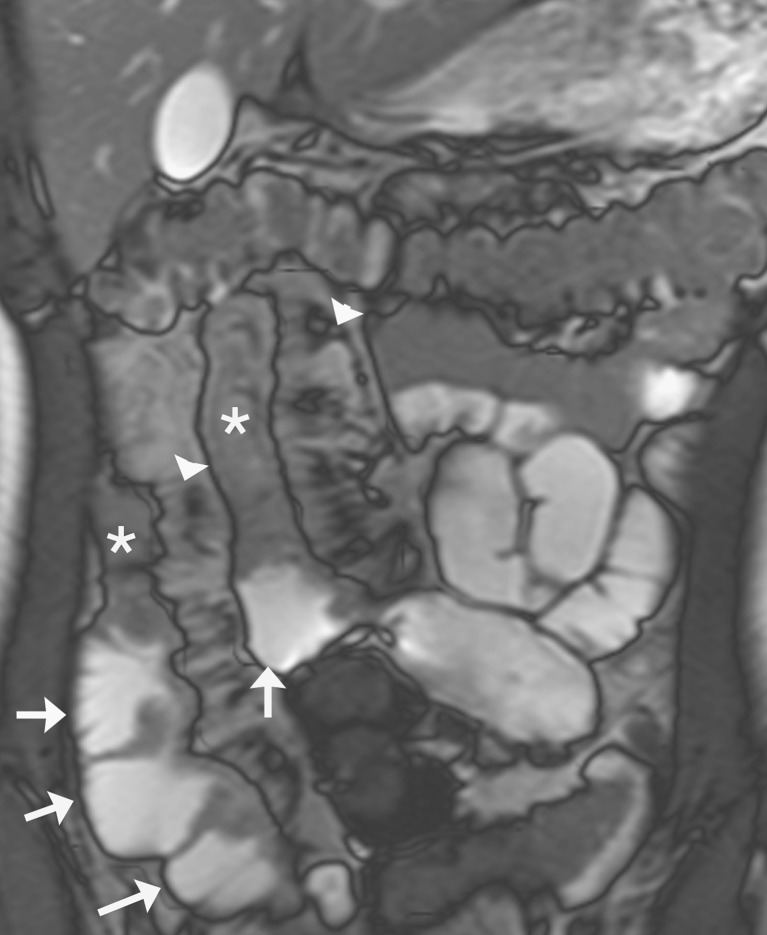
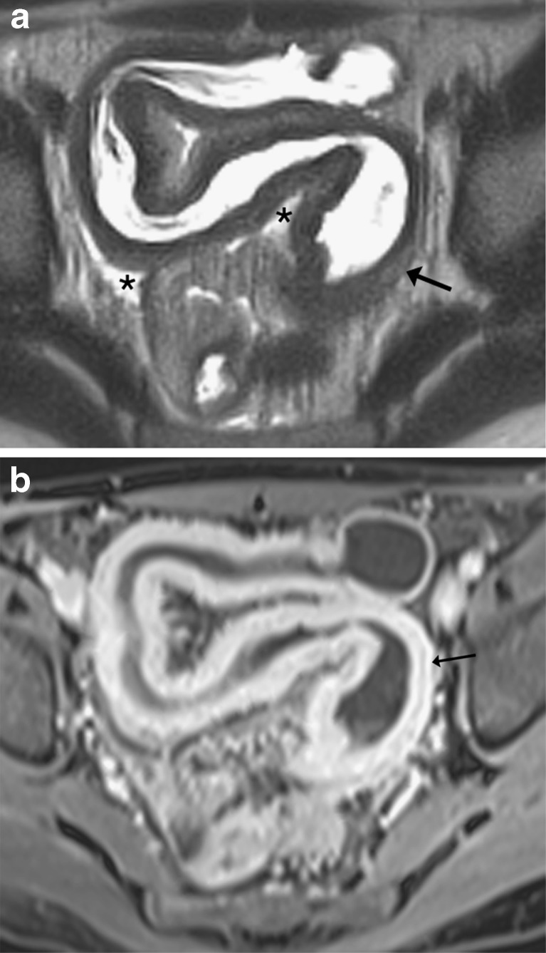
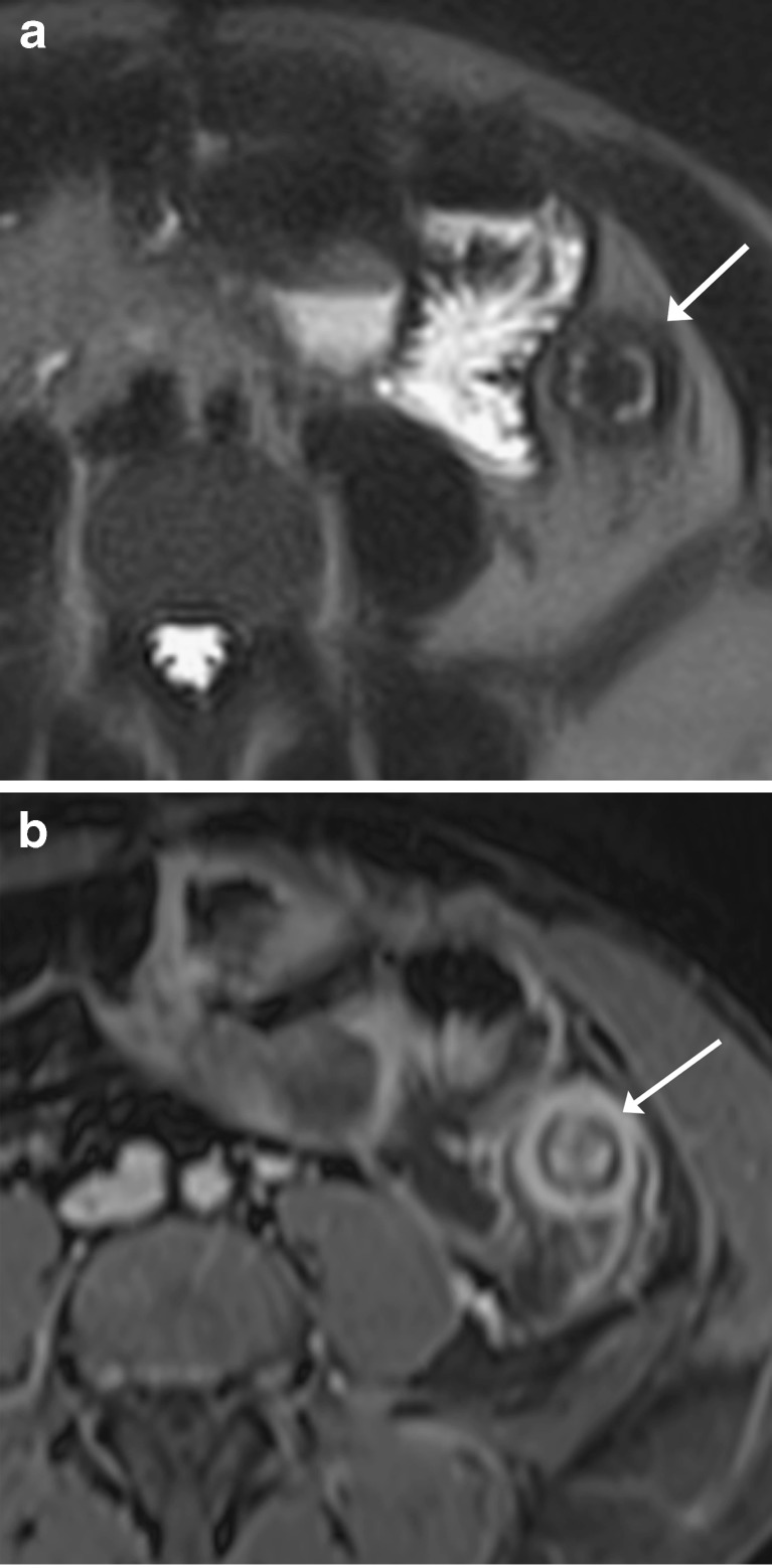
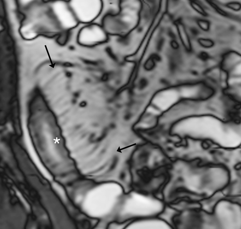
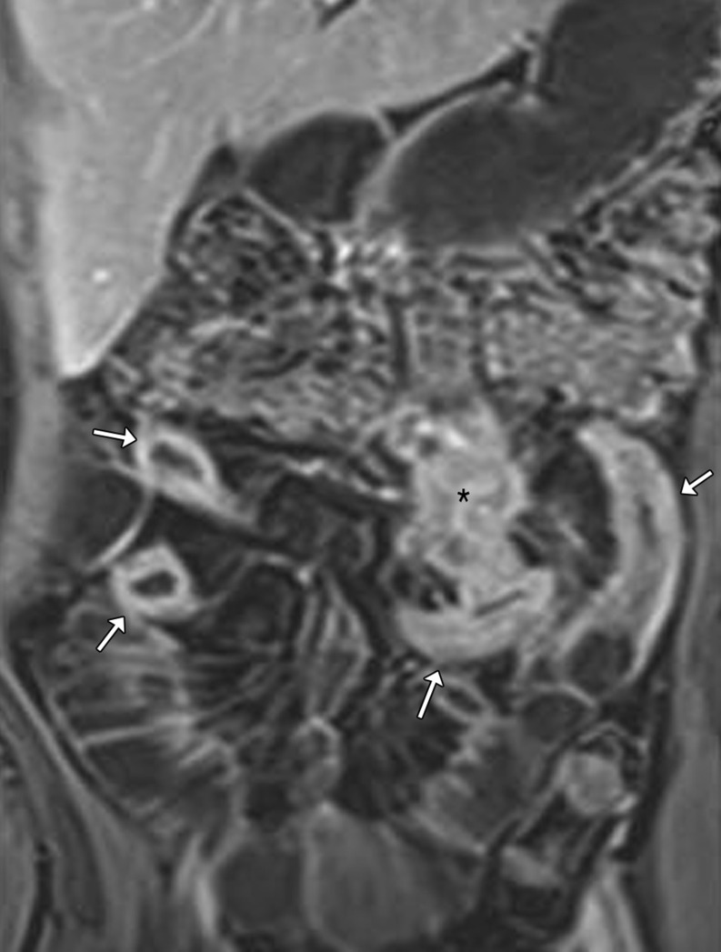
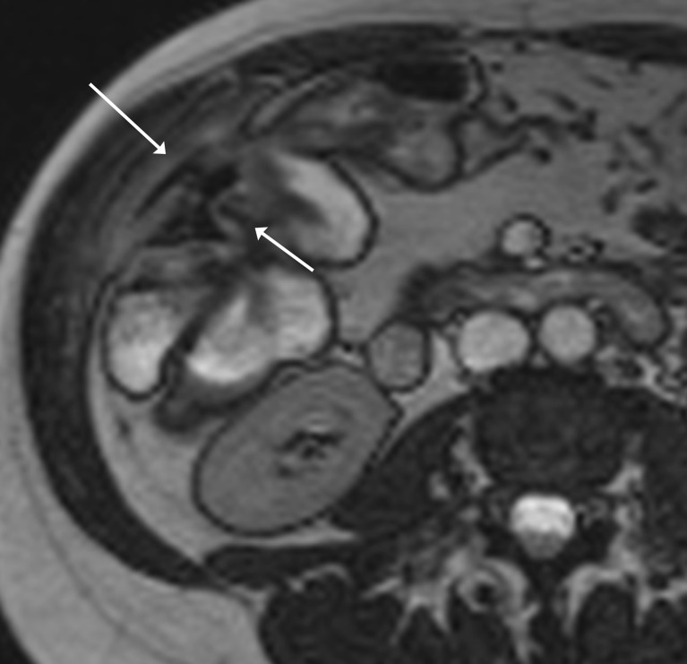
Magnetic resonance enterography (MRE) is fast becoming the first-line radiological investigation to evaluate the small bowel in patients with Crohn's disease. It can demonstrate both mural and extramural complications. The lack of ionizing radiation, together with high-contrast resolution, multiplanar capability and cine-imaging make it an attractive imaging modality in such patients who need prolonged follow-up. A key question in the management of such patients is the assessment of disease activity. Clinical indices, endoscopic and histological findings have traditionally been used as surrogate markers but all have limitations. MRE can help address this question. The purpose of this pictorial review is to (1) detail the MRE protocol used at our institution; (2) describe the rationale for the MR sequences used and their limitations; (3) compare MRE with other small bowel imaging techniques; (4) discuss how MRE can help distinguish between inflammatory, stricturing and penetrating disease, and thus facilitate management of this difficult condition. Main Messages • MR enterography (MRE) is the preferred imaging investigation to assess Crohn's disease. T2-weighted, post-contrast and diffusion-weighted imaging (DWI) can be used. • MRE offers no radiation exposure, high-contrast resolution, multiplanar ability and cine imaging. • MRE can help define disease activity, a key question in the management of Crohn's disease. • MRE can help distinguish between inflammatory, stricturing and penetrating disease. • MRE can demonstrate both mural and extramural complications.
磁共振肠造影术(MRE)正迅速成为评估克罗恩病患者小肠的首选放射学检查方法。它可以显示壁内和壁外并发症。缺乏电离辐射,以及高对比度分辨率、多平面能力和电影成像,使它成为需要长期随访的此类患者的一种有吸引力的成像方式。在这类患者的管理中,一个关键问题是评估疾病活动。临床指标、内镜和组织学发现传统上被用作替代标志物,但都有其局限性。MRE 可以帮助解决这个问题。本影像学综述的目的是:(1)详细介绍我们机构使用的 MRE 方案;(2)描述用于磁共振序列的原理及其局限性;(3)比较 MRE 与其他小肠成像技术;(4)讨论 MRE 如何有助于区分炎症性、狭窄性和穿透性疾病,从而有助于这种困难疾病的管理。主要信息 • 磁共振肠造影术(MRE)是评估克罗恩病的首选影像学检查方法。可使用 T2 加权、对比后和弥散加权成像(DWI)。 • MRE 不产生辐射,具有高对比度分辨率、多平面能力和电影成像。 • MRE 有助于定义疾病活动,这是克罗恩病管理中的一个关键问题。 • MRE 有助于区分炎症性、狭窄性和穿透性疾病。 • MRE 可以显示壁内和壁外并发症。