Judge Daniel P, Neamatalla Hany, Norris Russell A, Levine Robert A, Butcher Jonathan T, Vignier Nicolas, Kang Kevin H, Nguyen Quangtung, Bruneval Patrick, Perier Marie-Cécile, Messas Emmanuel, Jeunemaitre Xavier, de Vlaming Annemarieke, Markwald Roger, Carrier Lucie, Hagège Albert A
Faculté de Médecine, Université Paris Descartes, Sorbonne Paris Cité, Paris 75005 France; Division of Cardiology, Johns Hopkins University, Baltimore, MD 21287, USA.
Paris Cardiovascular Research Center, INSERM U970, Paris 75016 France; Department of Cardiology, Assistance Publique-Hôpitaux de Paris, Hôpital Européen Georges Pompidou, Paris 75016 France.
J Cardiovasc Dev Dis. 2015;2(2):48-65. doi: 10.3390/jcdd2020048. Epub 2015 Apr 21.
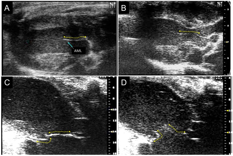
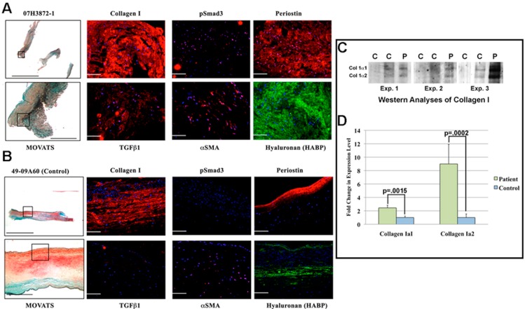
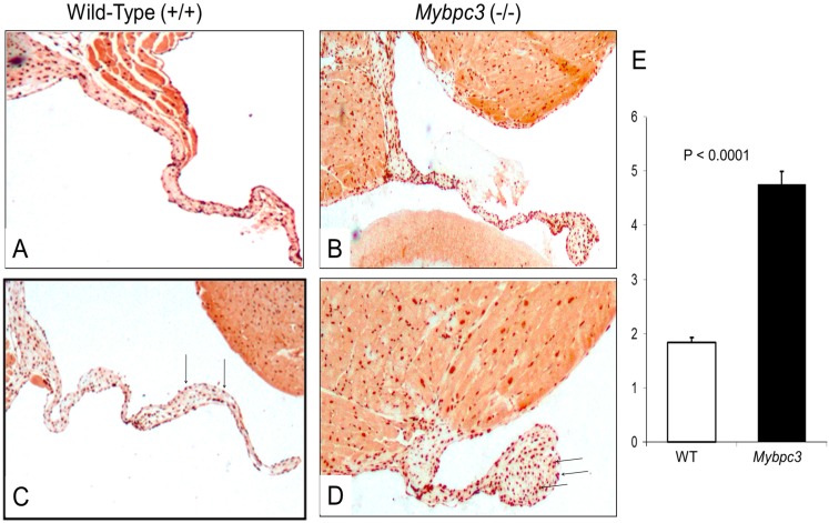
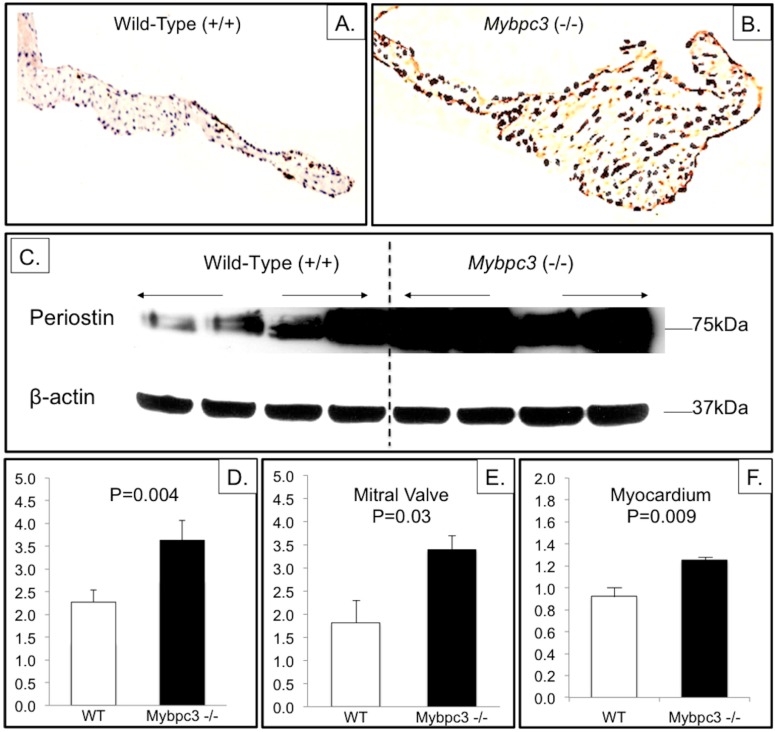
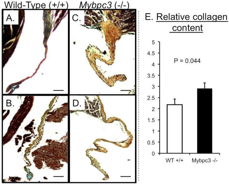
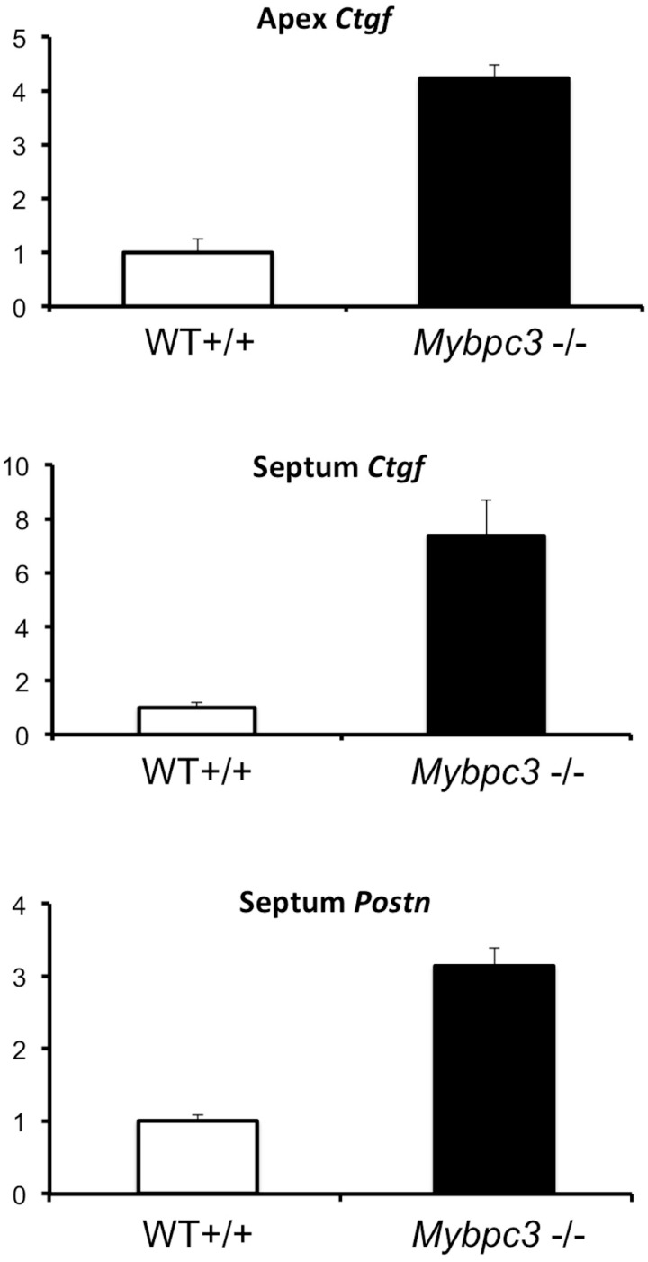
mutations cause hypertrophic cardiomyopathy, which is frequently associated with mitral valve (MV) pathology. We reasoned that increased MV size is caused by localized growth factors with paracrine effects. We used high-resolution echocardiography to compare -null, heterozygous, and wild-type mice ( = 84, aged 3-6 months) and micro-CT for MV volume ( = 6, age 6 months). -null mice showed left ventricular hypertrophy, dilation, and systolic dysfunction compared to heterozygous and wild-type mice, but no systolic anterior motion of the MV or left ventricular outflow obstruction. Compared to wild-type mice, echocardiographic anterior leaflet length (adjusted for left ventricular size) was greatest in -null mice (1.92 ± 0.08 . 1.72 ± 0.08 mm, < 0.001), as was combined leaflet thickness (0.23 ± 0.04 . 0.15 ± 0.02 mm, < 0.001). Micro-CT analyses of -null mice demonstrated increased MV volume (0.47 ± 0.06 . 0.15 ± 0.06 mm, = 0.018) and thickness (0.35 ± 0.04 . 0.12 ± 0.04 mm, = 0.002), coincident with increased markers of TGFβ activity compared to heterozygous and wild-type littermates. Similarly, excised MV from a patient with mutation showed increased TGFβ activity. We conclude that MYBPC3 deficiency causes hypertrophic cardiomyopathy with increased MV leaflet length and thickness despite the absence of left ventricular outflow-tract obstruction, in parallel with increased TGFβ activity. MV changes in hypertrophic cardiomyopathy may be due to paracrine effects, which represent targets for therapeutic studies.
突变会导致肥厚型心肌病,该病常与二尖瓣(MV)病变相关。我们推测MV大小增加是由具有旁分泌作用的局部生长因子引起的。我们使用高分辨率超声心动图比较了基因敲除、杂合子和野生型小鼠(每组n = 84只,年龄3 - 6个月),并使用微型计算机断层扫描(micro-CT)测量MV体积(每组n = 6只,年龄6个月)。与杂合子和野生型小鼠相比,基因敲除小鼠表现出左心室肥厚、扩张和收缩功能障碍,但没有MV收缩期前向运动或左心室流出道梗阻。与野生型小鼠相比,基因敲除小鼠超声心动图测量的前叶长度(根据左心室大小调整)最大(1.92±0.08 mm对1.72±0.08 mm,P < 0.001),联合叶厚度也是如此(0.23±0.04 mm对0.15±0.02 mm,P < 0.001)。对基因敲除小鼠的微型计算机断层扫描分析显示MV体积增加(0.47±0.06 mm对0.15±0.06 mm,P = 0.018)和厚度增加(0.35±0.04 mm对0.12±0.04 mm,P = 0.002),与杂合子和野生型同窝小鼠相比,TGFβ活性标志物增加。同样,来自一名携带该突变患者的切除MV显示TGFβ活性增加。我们得出结论,尽管没有左心室流出道梗阻,但MYBPC3缺乏会导致肥厚型心肌病,伴有MV叶长度和厚度增加,同时TGFβ活性增加。肥厚型心肌病中MV的变化可能是由于旁分泌作用,这代表了治疗研究的靶点。