Chen Aiqing, Oakley Arthur E, Monteiro Maria, Tuomela Katri, Allan Louise M, Mukaetova-Ladinska Elizabeta B, O'Brien John T, Kalaria Raj N
Neurovascular Research Group, Institute of Neuroscience, Newcastle University, Campus for Ageing & Vitality, Newcastle Upon Tyne, UK; Department of Psychiatry, University of Cambridge School of Clinical Medicine, Cambridge Biomedical Campus, Cambridge, UK.
Neurovascular Research Group, Institute of Neuroscience, Newcastle University, Campus for Ageing & Vitality, Newcastle Upon Tyne, UK; Department of Psychiatry, University of Cambridge School of Clinical Medicine, Cambridge Biomedical Campus, Cambridge, UK.
Neurobiol Aging. 2016 Feb;38:56-67. doi: 10.1016/j.neurobiolaging.2015.10.021. Epub 2015 Oct 30.
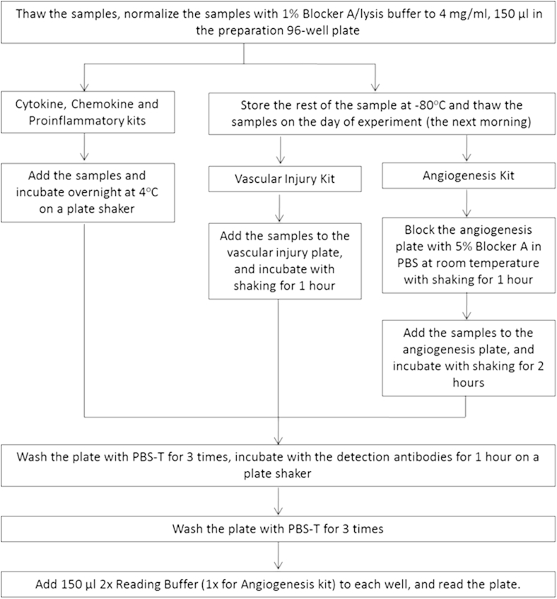
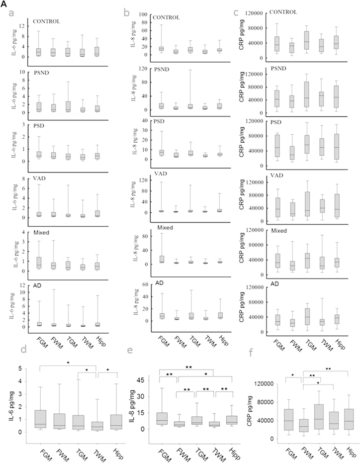
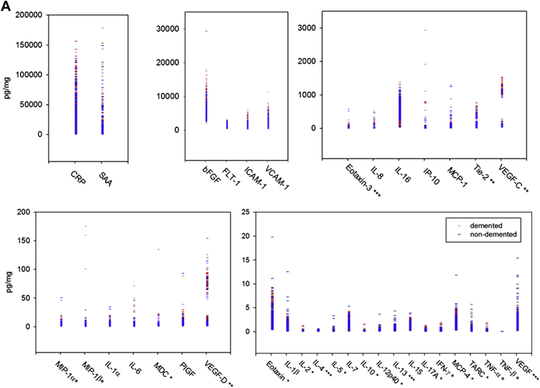
Both the inflammatory potential and cognitive function decline during aging. The association between the repertoire of inflammatory biomarkers and cognitive decline is unclear. Inflammatory cytokines have been reported to be increased, decreased, or unchanged in the cerebrospinal fluid and sera of subjects with dementia. We assessed 112 postmortem brains from subjects diagnosed with poststroke dementia (PSD), vascular dementia, mixed dementia, and Alzheimer's disease (AD), comparing those to poststroke nondemented (PSND) subjects and age-matched controls. We analyzed 5 brain regions including the gray and white matter from the frontal and temporal lobes for a panel of cytokine and/or chemokine analytes using multiplex-array assays. Of the 37 analytes, 14 were under or near the detection limits, 7 were close to the lowest detection level, and 16 cytokines were within the linear range of the assay. We observed widely variable concentrations of C-reactive protein (CRP) and serum amyloid A at the high end (1-150 ng/mg protein), whereas several of the interleukins (IL, interferon-gamma and tumor necrosis factor) at the low end (1-10 pg/mg). There were also regional variations; most notable being high concentrations of some cytokines (e.g., CRP and angiogenesis panel) in the frontal white matter. Overall, we found decreased concentrations of several cytokines, including IL-1 beta (p = 0.000), IL-6 (p = 0.000), IL-7 (p = 0.000), IL-8 (p = 0.000), IL-16 (p = 0.001), interferon-inducible protein-10 (0.044), serum amyloid A (p = 0.011), and a trend in IL-1 alpha (p = 0.084) across all dementia groups compared to nondemented controls. IL-6 and IL-8 were significantly lower in dementia subjects than in nondemented subjects in every region. In particular, lower levels of IL-6 and IL-8 were notable in the PSD compared to PSND subjects. Because these 2 stroke groups had comparable degree of vascular pathology, the lower production of IL-6 and IL-8 in PSD reaffirms a possible specific involvement of immunosenescence in dementia pathogenesis. In contrast, CRP was not altered between dementia and nondementia subjects or between PSD and PSND. Our study provides evidence not only for the feasibility of tracking cytokines in postmortem brain tissue but also suggests differentially impaired inflammatory mechanisms underlying dementia including AD. There was a diminished inflammatory response, possibly reflecting immunosenescence and cerebral atrophy, in all dementias. Strategies to enhance anti-inflammatory cytokines and boost the immune system of the brain may be beneficial for preventing cognitive dysfunction, especially after stroke.
炎症潜力和认知功能在衰老过程中都会下降。炎症生物标志物谱与认知衰退之间的关联尚不清楚。据报道,痴呆症患者脑脊液和血清中的炎症细胞因子水平升高、降低或无变化。我们评估了112例死后大脑,这些大脑来自被诊断为中风后痴呆(PSD)、血管性痴呆、混合性痴呆和阿尔茨海默病(AD)的患者,并将其与中风后无痴呆(PSND)患者及年龄匹配的对照组进行比较。我们使用多重阵列分析法,分析了包括额叶和颞叶灰质和白质在内的5个脑区中的一组细胞因子和/或趋化因子分析物。在37种分析物中,14种低于或接近检测限,7种接近最低检测水平,16种细胞因子在检测方法的线性范围内。我们观察到C反应蛋白(CRP)和血清淀粉样蛋白A在高端(1 - 150 ng/mg蛋白质)的浓度变化很大,而几种白细胞介素(IL、干扰素-γ和肿瘤坏死因子)在低端(1 - 10 pg/mg)。也存在区域差异;最显著的是额叶白质中一些细胞因子(如CRP和血管生成组)的浓度较高。总体而言,我们发现与非痴呆对照组相比,所有痴呆组中几种细胞因子的浓度降低,包括IL-1β(p = 0.000)、IL-6(p = 0.000)、IL-7(p = 0.000)、IL-8(p = 0.000)、IL-16(p = 0.001)、干扰素诱导蛋白-10(0.044)、血清淀粉样蛋白A(p = 0.011),并且IL-1α有下降趋势(p = 0.084)。在每个区域,痴呆症患者的IL-6和IL-8显著低于非痴呆患者。特别是,与PSND患者相比,PSD患者中IL-6和IL-8的水平更低。由于这两个中风组的血管病理学程度相当,PSD中IL-6和IL-8的产生较低再次证实了免疫衰老可能在痴呆症发病机制中具有特定作用。相比之下,痴呆症患者与非痴呆症患者之间或PSD与PSND之间CRP没有变化。我们的研究不仅为在死后脑组织中追踪细胞因子的可行性提供了证据,还表明包括AD在内的痴呆症潜在的炎症机制存在差异受损。在所有痴呆症中,炎症反应减弱,可能反映了免疫衰老和脑萎缩。增强抗炎细胞因子和增强大脑免疫系统的策略可能有助于预防认知功能障碍,尤其是在中风后。