Kangwantas Korakoch, Pinteaux Emmanuel, Penny Jeffrey
Manchester Pharmacy School, University of Manchester, Manchester, M13 9PT, UK.
Faculty of Life Sciences, University of Manchester, A.V. Hill Building, Oxford Road, Manchester, M13 9PT, UK.
J Neuroinflammation. 2016 Feb 1;13:25. doi: 10.1186/s12974-016-0495-9.
The blood-brain barrier (BBB) of the central nervous system (CNS) is essential for normal brain function. However, the loss of BBB integrity that occurs after ischaemic injury is associated with extracellular matrix (ECM) remodelling and inflammation, and contributes to poor outcome. ECM remodelling also contributes to BBB repair after injury, but the precise mechanisms and contribution of specific ECM molecules involved are unknown. Here, we investigated the mechanisms by which hypoxia and inflammation trigger loss of BBB integrity and tested the hypothesis ECM changes could contribute to BBB repair in vitro.
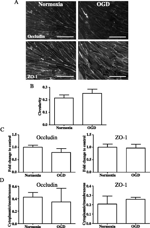
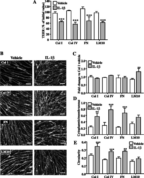
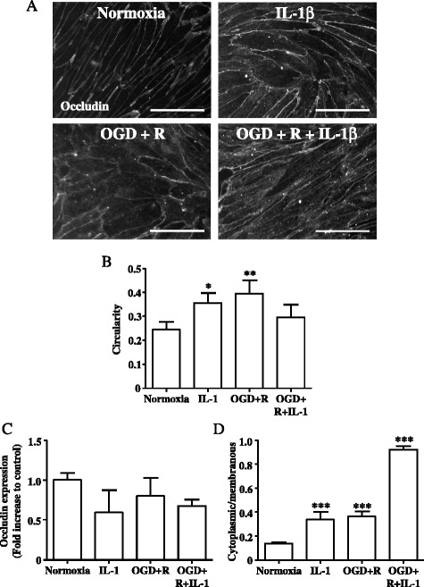
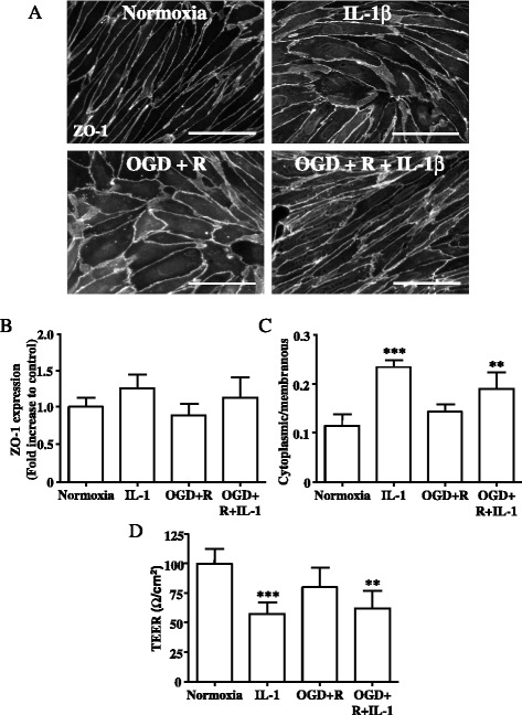
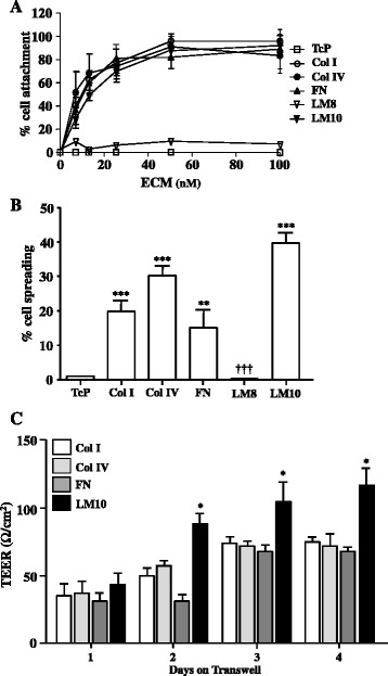
We used an in vitro model of the BBB, composed of primary rat brain endothelial cells grown on collagen (Col) I-, Col IV-, fibronectin (FN)-, laminin (LM) 8-, or LM10-coated tissue culture plates, either as a single monolayer culture or on Transwell® inserts above mixed glial cell cultures. Cultures were exposed to oxygen-glucose deprivation (OGD) and/or reoxygenation, in the absence or the presence of recombinant interleukin-1β (IL-1β). Cell adhesion to ECM molecules was assessed by cell attachment and cell spreading assays. BBB dysfunction was assessed by immunocytochemistry for tight junction proteins occludin and zona occludens-1 (ZO-1) and measurement of trans-endothelial electrical resistance (TEER). Change in endothelial expression of ECM molecules was assessed by semi-quantitative RT-PCR.
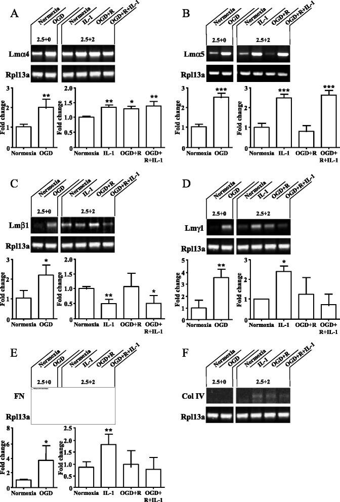
OGD and/or IL-1 induce dramatic changes associated with loss of BBB integrity, including cytoplasmic relocalisation of membrane-associated tight junction proteins occludin and ZO-1, cell swelling, and decreased TEER. OGD and IL-1 also induced gene expression of key ECM molecules associated with the BBB, including FN, Col IV, LM 8, and LM10. Importantly, we found that LM10, but not FN, Col IV, nor LM8, plays a key role in maintenance of BBB integrity and reversed most of the key hallmarks of BBB dysfunction induced by IL-1.
Our data unravel new mechanisms of BBB dysfunction induced by hypoxia and inflammation and identify LM10 as a key ECM molecule involved in BBB repair after hypoxic injury and inflammation.
中枢神经系统(CNS)的血脑屏障(BBB)对正常脑功能至关重要。然而,缺血性损伤后血脑屏障完整性的丧失与细胞外基质(ECM)重塑和炎症相关,并导致不良预后。ECM重塑也有助于损伤后血脑屏障的修复,但具体机制以及所涉及的特定ECM分子的作用尚不清楚。在此,我们研究了缺氧和炎症引发血脑屏障完整性丧失的机制,并测试了ECM变化有助于体外血脑屏障修复的假说。
我们使用了一种血脑屏障的体外模型,该模型由生长在I型胶原(Col)、IV型胶原(Col IV)、纤连蛋白(FN)、层粘连蛋白8(LM 8)或层粘连蛋白10(LM10)包被的组织培养板上的原代大鼠脑内皮细胞组成,可作为单层培养或置于混合神经胶质细胞培养物上方的Transwell®小室中。培养物在有无重组白细胞介素-1β(IL-1β)的情况下,暴露于氧葡萄糖剥夺(OGD)和/或复氧环境。通过细胞黏附和细胞铺展试验评估细胞对ECM分子的黏附。通过免疫细胞化学检测紧密连接蛋白闭合蛋白和封闭小带-1(ZO-1)以及测量跨内皮电阻(TEER)来评估血脑屏障功能障碍。通过半定量逆转录聚合酶链反应(RT-PCR)评估ECM分子在内皮细胞中的表达变化。
OGD和/或IL-1诱导了与血脑屏障完整性丧失相关的显著变化,包括膜相关紧密连接蛋白闭合蛋白和ZO-1的细胞质重新定位、细胞肿胀以及TEER降低。OGD和IL-1还诱导了与血脑屏障相关的关键ECM分子的基因表达,包括FN、Col IV、LM 8和LM10。重要的是,我们发现LM10而非FN、Col IV或LM8在维持血脑屏障完整性中起关键作用,并逆转了IL-1诱导的血脑屏障功能障碍的大多数关键特征。
我们的数据揭示了缺氧和炎症诱导血脑屏障功能障碍的新机制,并确定LM
10是缺氧损伤和炎症后参与血脑屏障修复的关键ECM分子。