Kim Woojun, Lee Jee Eun, Kim Su Hyun, Huh So Young, Hyun Jae Won, Jeong In Hye, Park Min Su, Cho Joong Yang, Lee Sang Hyun, Lee Kwang Soo, Kim Ho Jin
Department of Neurology, The Catholic University of Korea College of Medicine, Seoul, Korea.
Department of Neurology, Research Institute and Hospital of National Cancer Center, Goyang, Korea.
J Clin Neurol. 2016 Apr;12(2):188-93. doi: 10.3988/jcn.2016.12.2.188. Epub 2016 Jan 28.
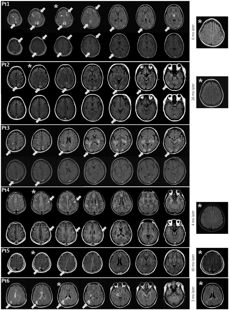
Brain lesions involving the cerebral cortex are rarely described in patients with neuromyelitis optica spectrum disorder (NMOSD), in contrast to multiple sclerosis. We investigated cerebral cortex involvement using conventional brain magnetic resonance imaging (MRI) in anti-aquaporin-4 (AQP4)-antibody-positive NMOSD patients.
The study enrolled 215 NMOSD patients who were seropositive for the anti-AQP4 antibody from 5 referral hospitals, and retrospectively analyzed their demographic, clinical, and MRI findings. Abnormal cerebral cortex lesions on brain MRI were identified by a neuroradiologist and two neurologists using consensus.
Most of the 215 enrolled patients (87%) were female. The median age at onset was 22.5 years (range: 15-36 years) and the mean follow-up duration was 123 months. Brain lesions were found in 143 of 194 patients (74%) in whom MRI was performed during follow-up. Brain lesions involving the cerebral cortex were identified in 6 of these 194 patients (3.1%). Five of the patients were female, and the six patients together had a median age of 29 years (range: 15-36 years) at the time of lesion presentation. Three of them showed leptomeningeal enhancement in the lesions. At presentation of the cortex-involving lesions, five of these patients were not being treated at the time of presentation, while the sixth was being treated with interferon-beta.
Although rare, cortical involvement occurs in NMOSD and is commonly combined with leptomeningeal enhancement. We speculate that this occurs only in patients who are not treated appropriately with immunosuppressant drugs.
与多发性硬化症不同,视神经脊髓炎谱系障碍(NMOSD)患者中很少描述涉及大脑皮层的脑损伤。我们使用传统脑磁共振成像(MRI)研究了抗水通道蛋白4(AQP4)抗体阳性的NMOSD患者的大脑皮层受累情况。
该研究纳入了来自5家转诊医院的215例抗AQP4抗体血清阳性的NMOSD患者,并回顾性分析了他们的人口统计学、临床和MRI检查结果。由一名神经放射科医生和两名神经科医生通过共识确定脑MRI上的大脑皮层异常病变。
纳入的215例患者中大多数(87%)为女性。发病的中位年龄为22.5岁(范围:15 - 36岁),平均随访时间为123个月。在随访期间进行MRI检查的194例患者中有143例(74%)发现有脑损伤。在这194例患者中有6例(3.1%)发现有涉及大脑皮层的脑损伤。其中5例患者为女性,这6例患者在病变出现时的中位年龄为29岁(范围:15 - 36岁)。其中3例病变处有软脑膜强化。在出现涉及大脑皮层的病变时,这些患者中有5例在病变出现时未接受治疗,而第6例正在接受β干扰素治疗。
虽然罕见,但NMOSD中会出现皮层受累,且通常合并软脑膜强化。我们推测这种情况仅发生在未接受免疫抑制药物适当治疗的患者中。