Lee Ji Young, Chung Hyewon, Kim Hyung Chan
Department of Ophthalmology, Konkuk University Medical Center, Konkuk University School of Medicine, Seoul, Korea.
Korean J Ophthalmol. 2016 Feb;30(1):17-24. doi: 10.3341/kjo.2016.30.1.17. Epub 2016 Jan 21.
To describe the changes of fundus autofluorescence (FAF) in patients with age-related macular degeneration before and after intravitreal injection of anti-vascular endothelial growth factor according to the type of choroidal neovascularization (CNV) and to evaluate the correlation of FAF with spectral domain optical coherence tomography (SD-OCT) parameters and vision.
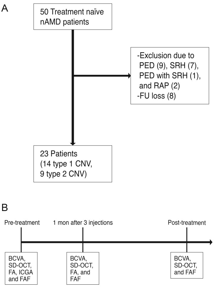
This was a retrospective study. Twenty-one treatment-naïve patients with neovascular age-related macular degeneration were included. Study eyes were divided into two groups according to the type of CNV. Fourteen eyes were type 1 CNV and seven eyes were type 2 CNV. All eyes underwent a complete ophthalmologic examination, including an assessment of best-corrected visual acuity, SD-OCT, fluorescein angiography, and FAF imaging, before and 3 months after intravitreal anti-vascular endothelial growth factor injection. Gray scales of FAF image for CNV areas, delineated as in fluorescein angiography, were analyzed using the ImageJ program, which were adjusted by comparison with normal background areas. Correlation of changes in FAF with changes in SD-OCT parameters, including CNV thickness, photoreceptor inner and outer segment junction disruption length, external limiting membrane disruption length, central macular thickness, subretinal fluid, and intraretinal fluid were analyzed.
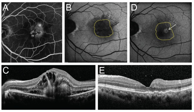
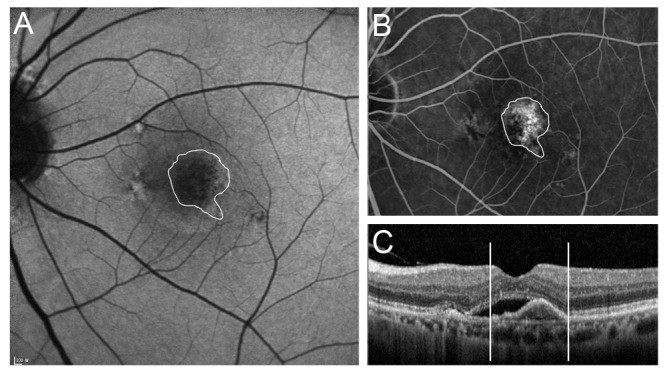
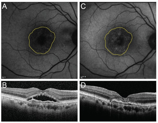
Eyes with both type 1 and type 2 CNV showed reduced FAF before treatment. The mean gray scales (%) of type 1 and type 2 CNV were 52.20% and 42.55%, respectively. The background values were 106.72 and 96.86. After treatment, the mean gray scales (%) of type 1 CNV and type 2 CNV were changed to 57.61% (p = 0.005) and 57.93% (p = 0.008), respectively. After treatment, CNV thickness, central macular thickness, and inner and outer segment junction disruption length were decreased while FAF increased.
FAF was noted to be reduced in eyes with newly diagnosed wet age-related macular degeneration, but increased after anti-vascular endothelial growth factor therapy regardless of CNV lesion type.
根据脉络膜新生血管(CNV)类型描述年龄相关性黄斑变性患者玻璃体内注射抗血管内皮生长因子前后的眼底自发荧光(FAF)变化,并评估FAF与光谱域光学相干断层扫描(SD-OCT)参数及视力的相关性。
这是一项回顾性研究。纳入21例初治的新生血管性年龄相关性黄斑变性患者。研究眼根据CNV类型分为两组。14只眼为1型CNV,7只眼为2型CNV。所有眼在玻璃体内注射抗血管内皮生长因子前及注射后3个月均接受了全面的眼科检查,包括最佳矫正视力评估、SD-OCT、荧光素血管造影和FAF成像。使用ImageJ程序分析荧光素血管造影中划定的CNV区域的FAF图像灰度值,并与正常背景区域比较进行调整。分析FAF变化与SD-OCT参数变化的相关性,这些参数包括CNV厚度、光感受器内外节连接破坏长度、外界膜破坏长度、黄斑中心厚度、视网膜下液和视网膜内液。
1型和2型CNV的眼在治疗前FAF均降低。1型和2型CNV的平均灰度值(%)分别为52.20%和42.55%。背景值分别为106.72和96.86。治疗后,1型CNV和2型CNV的平均灰度值(%)分别变为57.61%(p = 0.005)和57.93%(p = 0.008)。治疗后,CNV厚度、黄斑中心厚度以及内外节连接破坏长度减小,而FAF增加。
新诊断的湿性年龄相关性黄斑变性患者的眼FAF降低,但抗血管内皮生长因子治疗后FAF增加,且与CNV病变类型无关。