University of Ottawa, Ottawa, Ontario, Canada.
University of Leeds and NIHR Leeds Musculoskeletal Biomedical Research Unit, Leeds, UK.
Arthritis Rheumatol. 2016 Jul;68(7):1648-59. doi: 10.1002/art.39622.
In patients with osteoarthritis (OA), bone marrow lesions (BMLs) are intimately linked to disease progression. We hypothesized that aberrant multipotential stromal cell (also known as mesenchymal stem cell [MSC]) responses within bone tissue contributes to BML pathophysiology. The aim of this study was to investigate BML and non-BML native subchondral bone MSCs for numeric, topographic, in vitro functional, and gene expression differences.
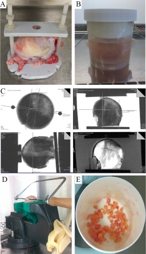
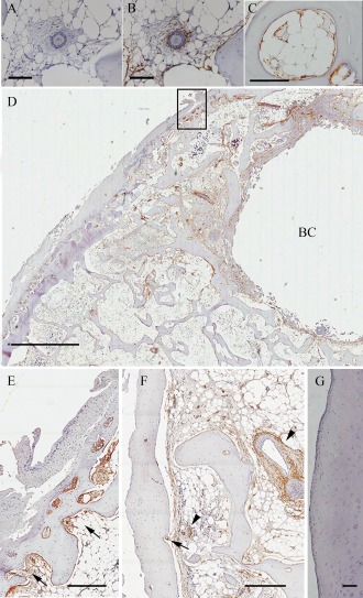
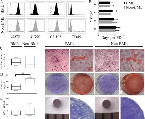
Ex vivo 3T magnetic resonance imaging (MRI) of the femoral heads of 20 patients with hip OA was performed. MRI-determined BML and non-BML regions were excised and enzymatically treated to extract cells and quantify MSCs using flow cytometry and colony-forming unit-fibroblast (CFU-F) assay. Immunohistochemical analysis was performed to determine in vivo CD271+ MSC distribution. Culture-expanded CD271+ cells were analyzed for tripotentiality and gene expression.
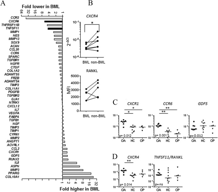
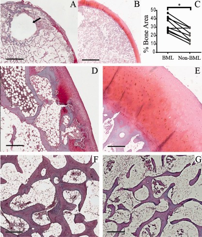
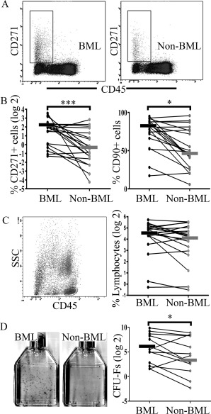
BML regions were associated with greater trabecular bone area and cartilage damage compared with non-BML regions. The proportion of CD45-CD271+ MSCs was higher in BML regions compared with non-BML regions (median difference 5.6-fold; P < 0.001); the CFU-F assay showed a similar trend (median difference 4.3-fold; P = 0.013). Immunohistochemistry revealed CD271+ cell accumulation in bone adjacent to cartilage defects and areas of osteochondral angiogenesis. BML MSCs had lower proliferation and mineralization capacities in vitro and altered expression of TNFSF11/RANKL and CXCR4/stromal cell-derived factor 1 receptor. OA MSCs showed up-regulated transcripts for CXCR1 and CCR6 compared with MSCs derived from healthy or osteoporotic bone.
This study is the first to show numeric and topographic alterations in native MSCs in the diseased bone of patients with hip OA. Given the associated functional perturbation of MSCs, these data suggest that subchondral bone MSC manipulation may be an OA treatment target.
在骨关节炎(OA)患者中,骨髓病变(BML)与疾病进展密切相关。我们假设骨组织内异常的多能基质细胞(也称为间充质干细胞[MSC])反应导致 BML 病理生理学发生。本研究旨在研究 BML 和非 BML 原发性软骨下骨 MSC 的数量、地形、体外功能和基因表达差异。
对 20 例髋 OA 患者的股骨头进行 3T 磁共振成像(MRI)的体外研究。通过 MRI 确定 BML 和非 BML 区域,并进行酶处理以提取细胞,并使用流式细胞术和集落形成单位-成纤维细胞(CFU-F)测定法对 MSC 进行定量。进行免疫组织化学分析以确定体内 CD271+MSC 分布。对培养扩增的 CD271+细胞进行三潜能性和基因表达分析。
BML 区域与非 BML 区域相比,小梁骨面积更大,软骨损伤更严重。与非 BML 区域相比,BML 区域的 CD45-CD271+MSC 比例更高(中位数差异 5.6 倍;P<0.001);CFU-F 测定也显示出类似的趋势(中位数差异 4.3 倍;P=0.013)。免疫组织化学显示 CD271+细胞在软骨缺损和骨软骨血管生成区域的软骨下骨中积聚。BML MSC 的体外增殖和矿化能力较低,并且 TNFSF11/RANKL 和 CXCR4/基质细胞衍生因子 1 受体的表达发生改变。与来自健康或骨质疏松骨的 MSC 相比,OA MSC 显示出 CXCR1 和 CCR6 的转录本上调。
本研究首次显示髋 OA 患者患病骨中天然 MSC 的数量和地形改变。鉴于 MSC 的相关功能紊乱,这些数据表明软骨下骨 MSC 操作可能是 OA 治疗的靶点。