Department of Biomedical Science, College of Life Science, CHA University, 6F, CHA bio-complex, 689 Sampyeong-Dong, Bundang-Gu, Seongnam-Si Republic of Korea.
Biomater Res. 2016 Feb 18;20:4. doi: 10.1186/s40824-016-0051-9. eCollection 2016.
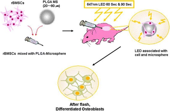
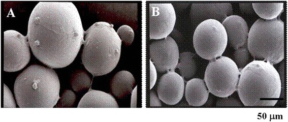
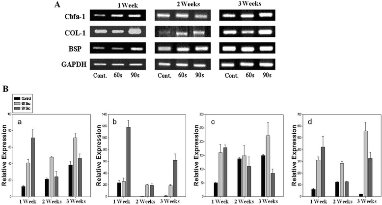
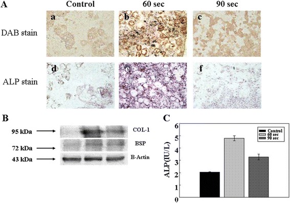
Biodegradable microspheres fabricated from poly (Lactic-co-glycolic acid) (PLGA) have attracted considerable attention in the bone tissue regeneration field. In this study, rabbit mesenchymal stem cells (rMSCs) adherent to PLGA microspheres were implanted into athymic nude mice and irradiated with 647 nm red light to promote bone formation. It was found that irradiating rMSCs with high levels of red light (647 nm) from an LED (light-emitting diode) increased levels of bone specific markers in rMSCs embedded on PLGA microspheres.
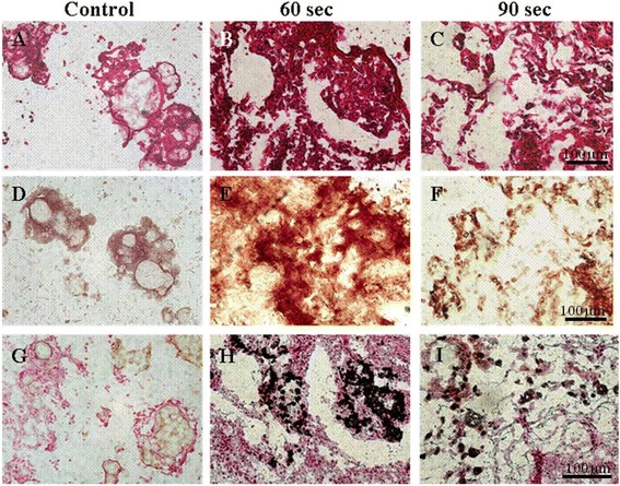
These increased expressions were observed by RT-PCR, real time-QPCR, immunohistochemistry (IHC), and von Kossa and Alizarin red S staining. Microsphere matrices coated with rMSCs were injected into athymic nude mice and irradiated with red light for 60 seconds showed significantly greater bone-specific phenotypes after 4 weeks in vivo.
The devised PLGA microsphere matrix containing rMSCs and irradiation with red light at 647 nm process shows promise as a means of coating implantable biomedical devices to improve their biocompatibilities and in vivo performances.
聚(乳酸-共-乙醇酸)(PLGA)制成的可生物降解微球在骨组织再生领域引起了相当大的关注。在这项研究中,将附着在 PLGA 微球上的兔间充质干细胞(rMSCs)植入无胸腺裸鼠,并辐照 647nm 红光以促进骨形成。结果发现,用 LED(发光二极管)发出的高剂量 647nm 红光辐照 rMSCs 会增加嵌入 PLGA 微球中的 rMSCs 的骨特异性标志物的水平。
通过 RT-PCR、实时 QPCR、免疫组织化学(IHC)和 von Kossa 及茜素红 S 染色观察到这些表达增加。将涂覆有 rMSCs 的微球基质注入无胸腺裸鼠,并辐照 60 秒红光,在体内 4 周后显示出明显更强的骨特异性表型。
设计的含有 rMSCs 的 PLGA 微球基质,并辐照 647nm 红光,显示出作为一种涂覆可植入生物医学设备的方法的潜力,以提高其生物相容性和体内性能。