Saunders Arpiar, Huang Kee Wui, Sabatini Bernardo Luis
Department of Neurobiology, Howard Hughes Medical Institute, Harvard Medical School, Boston, Massachusetts, United States of America.
PLoS One. 2016 Feb 23;11(2):e0149798. doi: 10.1371/journal.pone.0149798. eCollection 2016.
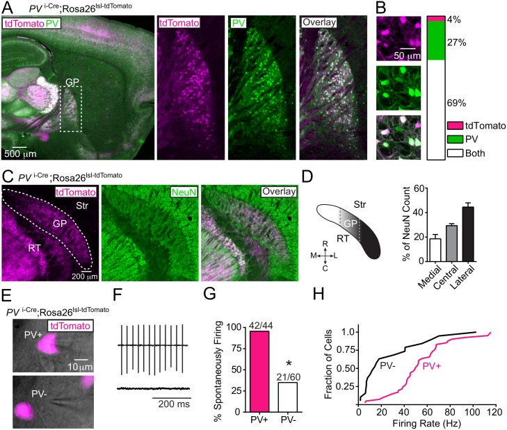
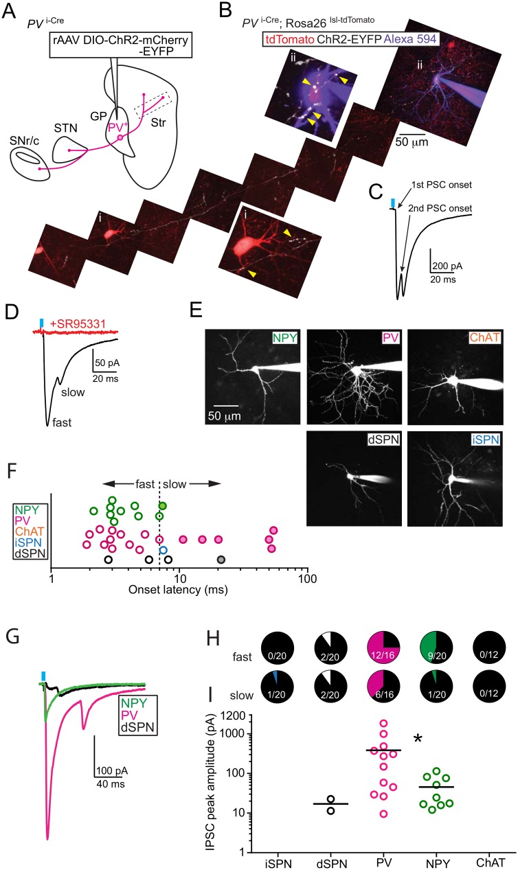
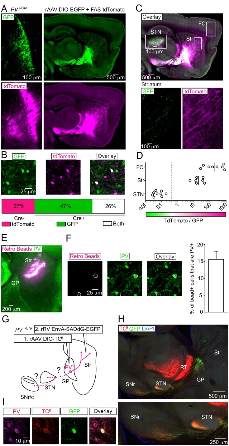
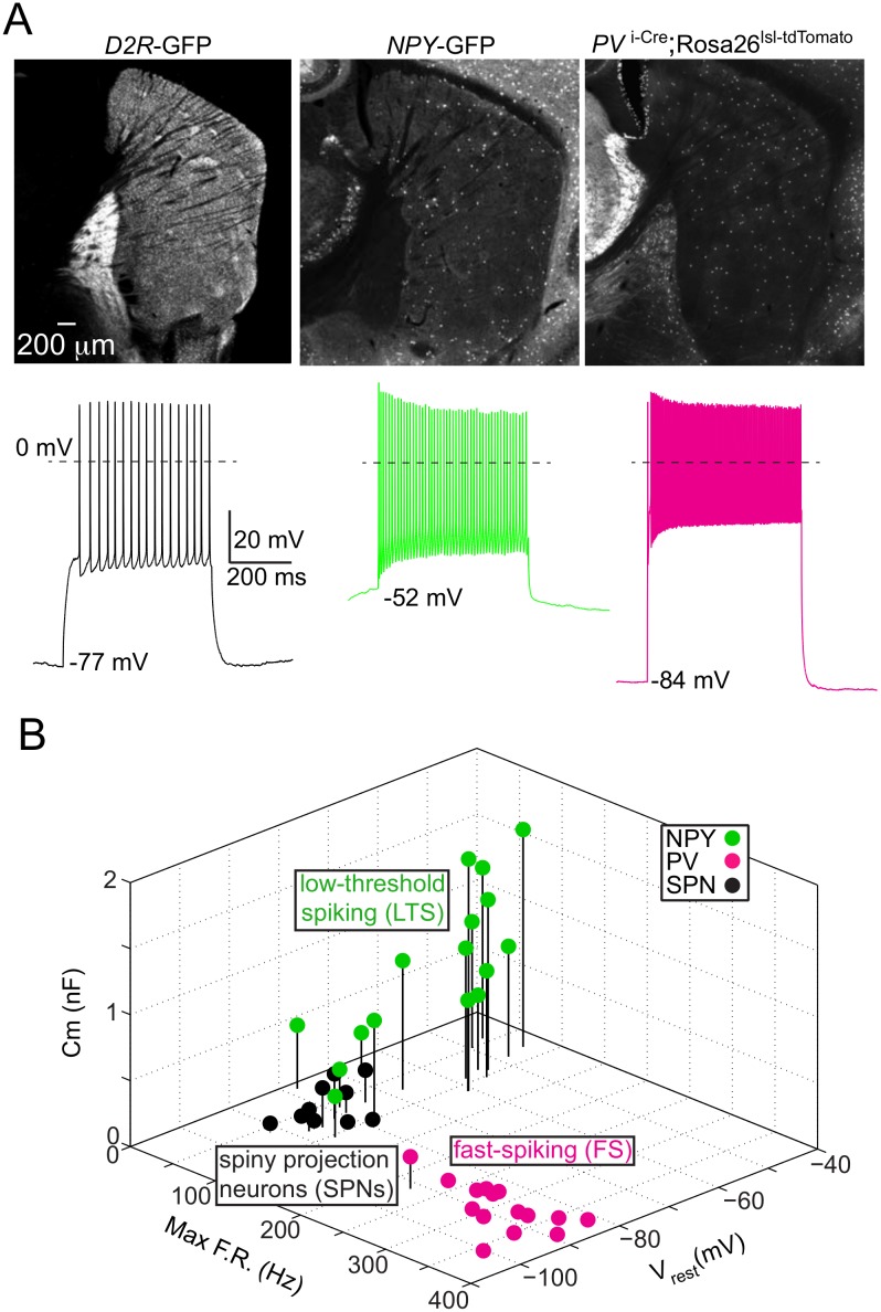
The globus pallidus externus (GP) is a nucleus of the basal ganglia (BG), containing GABAergic projection neurons that arborize widely throughout the BG, thalamus and cortex. Ongoing work seeks to map axonal projection patterns from GP cell types, as defined by their electrophysiological and molecular properties. Here we use transgenic mice and recombinant viruses to characterize parvalbumin expressing (PV+) GP neurons within the BG circuit. We confirm that PV+ neurons 1) make up ~40% of the GP neurons 2) exhibit fast-firing spontaneous activity and 3) provide the major axonal arborization to the STN and substantia nigra reticulata/compacta (SNr/c). PV+ neurons also innervate the striatum. Retrograde labeling identifies ~17% of pallidostriatal neurons as PV+, at least a subset of which also innervate the STN and SNr. Optogenetic experiments in acute brain slices demonstrate that the PV+ pallidostriatal axons make potent inhibitory synapses on low threshold spiking (LTS) and fast-spiking interneurons (FS) in the striatum, but rarely on spiny projection neurons (SPNs). Thus PV+ GP neurons are synaptically positioned to directly coordinate activity between BG input nuclei, the striatum and STN, and thalamic-output from the SNr.
苍白球外侧部(GP)是基底神经节(BG)的一个核团,包含γ-氨基丁酸能投射神经元,其树突广泛分布于整个基底神经节、丘脑和皮质。正在进行的研究试图描绘出由电生理和分子特性定义的GP细胞类型的轴突投射模式。在这里,我们使用转基因小鼠和重组病毒来表征基底神经节回路中表达小白蛋白(PV+)的GP神经元。我们证实,PV+神经元:1)占GP神经元的约40%;2)表现出快速放电的自发活动;3)向丘脑底核(STN)和黑质网状部/致密部(SNr/c)提供主要的轴突分支。PV+神经元也支配纹状体。逆行标记显示,约17%的苍白球-纹状体神经元为PV+,其中至少一部分也支配STN和SNr。急性脑片的光遗传学实验表明,PV+苍白球-纹状体轴突在纹状体的低阈值发放(LTS)和快速发放中间神经元(FS)上形成有效的抑制性突触,但很少在棘状投射神经元(SPN)上形成突触。因此,PV+ GP神经元在突触位置上可直接协调基底神经节输入核团、纹状体和STN之间的活动,以及SNr的丘脑输出。