Shukla Sanket Kumar, Jha Hem Chandra, El-Naccache Darine W, Robertson Erle S
Department of Otorhinolaryngology and Tumor Virology Program, Abramson Cancer Center, Perelman School of Medicine at The University of Pennsylvania, Philadelphia, PA-19104, USA.
Oncotarget. 2016 Apr 5;7(14):18116-34. doi: 10.18632/oncotarget.7502.
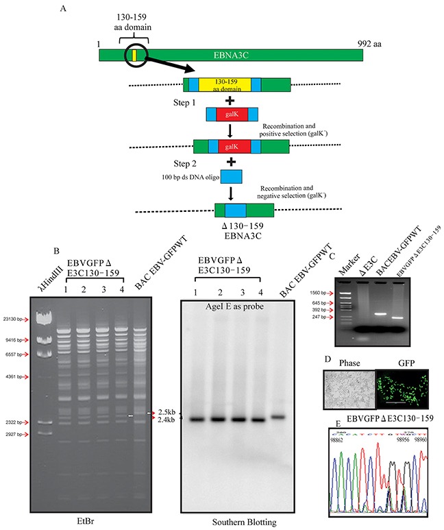
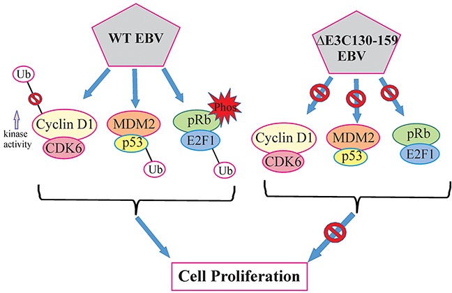
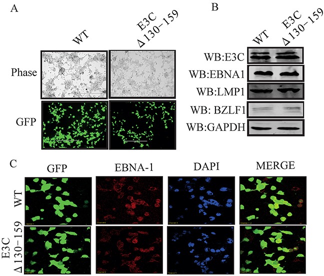
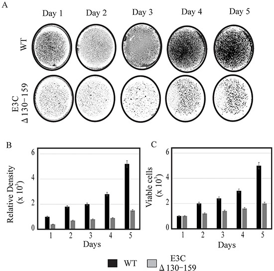
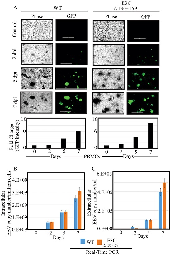
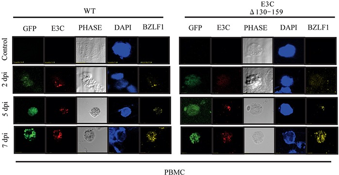
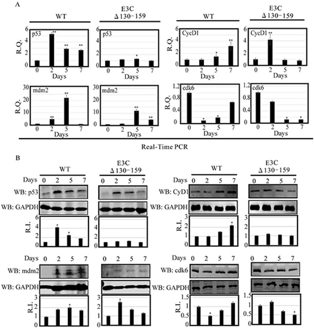
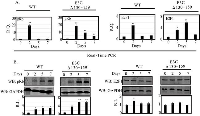
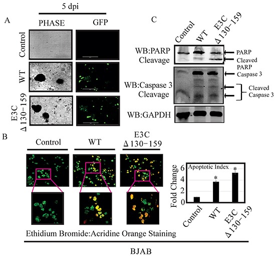
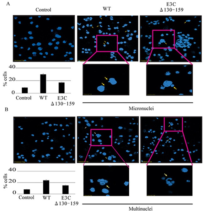
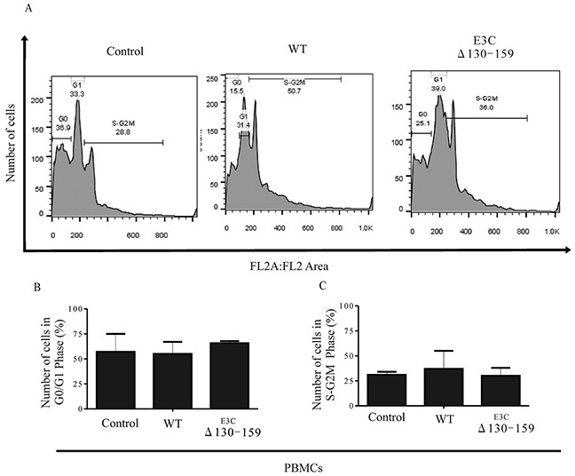
Epstein-Barr virus (EBV), a gamma herpes virus is associated with B-cell malignancies. EBNA-3C is critical for in vitro primary B-cell transformation. Interestingly, the N terminal domain of EBNA3C which contains residues 130-159, interacts with various cellular proteins, such as p53, Mdm2, CyclinD1/Cdk6 complex, and E2F1. In the current reverse genetics study, we deleted the residues 130-159 aa within EBNA3C open reading frame (ORF) by BACmid recombinant engineering methodology. Our experiments demonstrated that deletion of the 130-159 aa showed a reduction in cell proliferation. Also, this recombinant virus showed with higher infectivity of human peripheral blood mononuclear cells (PBMCs) compared to wild type EBV. PBMCs- infected with recombinant EBV deleted for 130-159 residues have differential expression patterns for the p53/Mdm2, CyclinD1/Cdk6 and pRb/E2F1 pathways compared to wild type EBV-infected PBMCs. PBMCs infected with recombinant virus showed increased apoptotic cell death which further resulted in activation of polymerase 1 (PARP1), an important contributor to apoptotic signaling. Interestingly, cells infected with this recombinant virus showed a dramatic decrease in chromosomal instability, indicated by the presence of increased multinucleation and micronucleation. In addition infection with recombinant virus have increased cells in G0/G1 phase and decreased cells in S-G2M phase when compared to wild type infected cells. Thus, these differences in signaling activities due to 29 amino acid residues of EBNA3C is of particular significance in deregulation of cell proliferation in EBV-infected cells.
爱泼斯坦-巴尔病毒(EBV)是一种γ疱疹病毒,与B细胞恶性肿瘤有关。EBNA-3C对体外原代B细胞转化至关重要。有趣的是,EBNA3C的N末端结构域(包含130-159位残基)与多种细胞蛋白相互作用,如p53、Mdm2、细胞周期蛋白D1/Cdk6复合物和E2F1。在当前的反向遗传学研究中,我们通过BACmid重组工程方法删除了EBNA3C开放阅读框(ORF)内的130-159位氨基酸残基。我们的实验表明,删除130-159位氨基酸残基会导致细胞增殖减少。此外,与野生型EBV相比,这种重组病毒对人外周血单核细胞(PBMC)具有更高的感染性。与野生型EBV感染的PBMC相比,感染了缺失130-159位残基的重组EBV的PBMC在p53/Mdm2、细胞周期蛋白D1/Cdk6和pRb/E2F1信号通路中具有不同的表达模式。感染重组病毒的PBMC显示凋亡细胞死亡增加,这进一步导致凋亡信号的重要贡献者聚合酶1(PARP1)的激活。有趣的是,感染这种重组病毒的细胞显示染色体不稳定性显著降低,表现为多核化和微核化增加。此外,与野生型感染细胞相比,感染重组病毒的细胞在G0/G1期增加,在S-G2M期减少。因此,EBNA3C的29个氨基酸残基导致的这些信号活性差异在EBV感染细胞的细胞增殖失调中具有特别重要的意义。