Jalbert Llewellyn E, Neill Evan, Phillips Joanna J, Lupo Janine M, Olson Marram P, Molinaro Annette M, Berger Mitchel S, Chang Susan M, Nelson Sarah J
Joint Graduate Program in Bioengineering (L.E.J., S.J.N.), Department of Radiology & Biomedical Imaging (E.N., J.M.L., M.P.O., S.J.N.), Department of Pathology (J.J.P.), Department of Neurological Surgery (J.J.P., A.M.M., M.S.B., S.M.C.), Department of Biostatistics and Epidemiology (A.M.M.), University of California, San Francisco, San Francisco, California.
Neuro Oncol. 2016 Aug;18(8):1169-79. doi: 10.1093/neuonc/now008. Epub 2016 Feb 23.
Patients with low-grade glioma (LGG) have a relatively long survival, and a balance is often struck between treating the tumor and impacting quality of life. While lesions may remain stable for many years, they may also undergo malignant transformation (MT) at the time of recurrence and require more aggressive intervention. Here we report on a state-of-the-art multiparametric MRI study of patients with recurrent LGG.
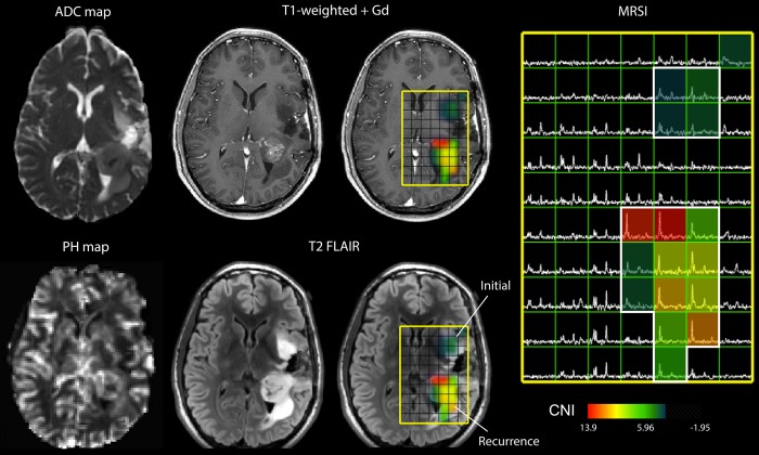
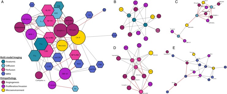
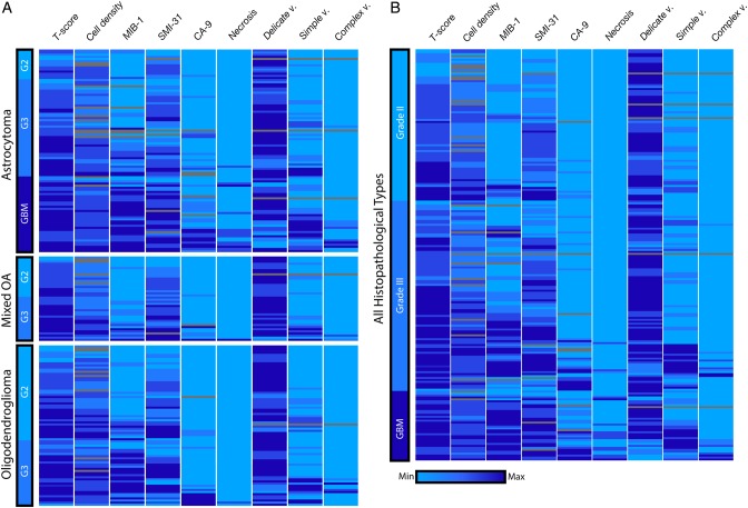
One hundred and eleven patients previously diagnosed with LGG were scanned at either 1.5 T or 3 T MR at the time of recurrence. Volumetric and intensity parameters were estimated from anatomic, diffusion, perfusion, and metabolic MR data. Direct comparisons of histopathological markers from image-guided tissue samples with metrics derived from the corresponding locations on the in vivo images were made. A bioinformatics approach was applied to visualize and interpret these results, which included imaging heatmaps and network analysis. Multivariate linear-regression modeling was utilized for predicting transformation.
Many advanced imaging parameters were found to be significantly different for patients with tumors that had undergone MT versus those that had not. Imaging metrics calculated at the tissue sample locations highlighted the distinct biological significance of the imaging and the heterogeneity present in recurrent LGG, while multivariate modeling yielded a 76.04% accuracy in predicting MT.
The acquisition and quantitative analysis of such multiparametric MR data may ultimately allow for improved clinical assessment and treatment stratification for patients with recurrent LGG.
低级别胶质瘤(LGG)患者生存期相对较长,治疗肿瘤与影响生活质量之间往往需要权衡。虽然病灶可能多年保持稳定,但复发时也可能发生恶性转化(MT),需要更积极的干预。在此,我们报告一项关于复发性LGG患者的先进多参数MRI研究。
111例先前诊断为LGG的患者在复发时接受了1.5T或3T MR扫描。从解剖、扩散、灌注和代谢MR数据中估计体积和强度参数。对图像引导组织样本的组织病理学标志物与体内图像相应位置得出的指标进行直接比较。应用生物信息学方法可视化和解释这些结果,包括成像热图和网络分析。利用多元线性回归模型预测转化。
发现许多先进成像参数在发生MT的肿瘤患者与未发生MT的患者之间存在显著差异。在组织样本位置计算的成像指标突出了成像的独特生物学意义以及复发性LGG中存在的异质性,而多元建模在预测MT方面的准确率为76.04%。
此类多参数MR数据的采集和定量分析最终可能有助于改善复发性LGG患者的临床评估和治疗分层。