Gargiulo Paolo, Helgason Thordur, Ramon Ceon, Jónsson Halldór, Carraro Ugo
CIR-Myo, Translational Myology Lab, Department of Biomedical Sciences, University of Padova , Italy.
Eur J Transl Myol. 2014 Mar 27;24(1):3298. doi: 10.4081/ejtm.2014.3298. eCollection 2014 Mar 31.
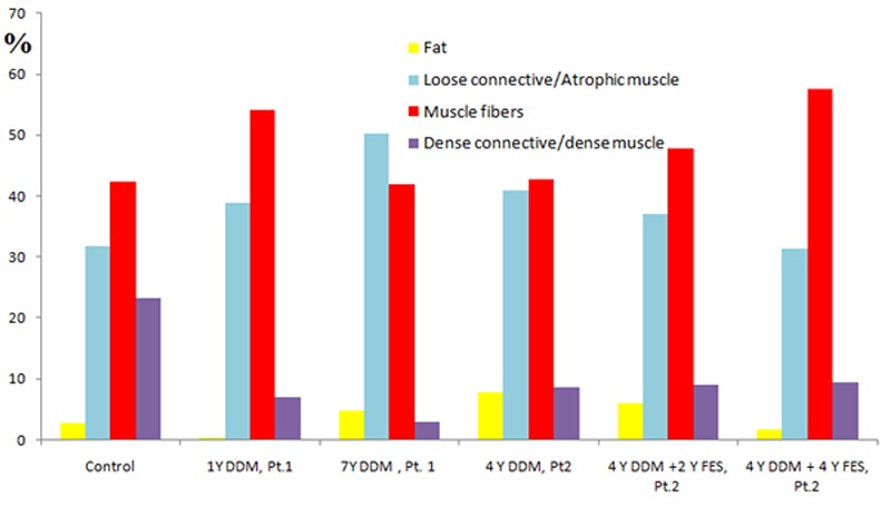
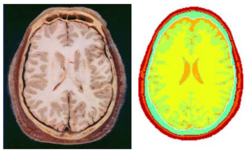
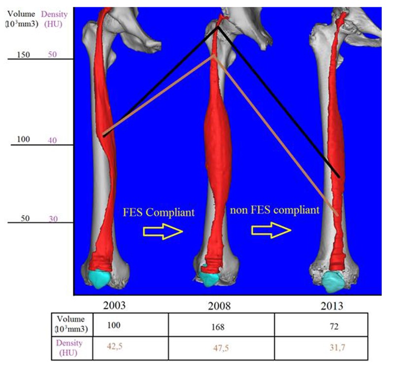
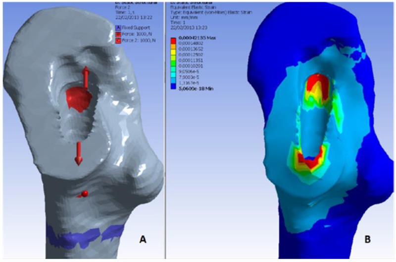
This paper reviews the novel use of CT and MRI data and image processing tools to segment and reconstruct tissue images in 3D to determine characteristics of muscle, bone and brain. This to study and simulate the structural changes occurring in healthy and pathological conditions as well as in response to clinical treatments. Here we report the application of this methodology to evaluate and quantify: 1. progression of atrophy in human muscle subsequent to permanent lower motor neuron (LMN) denervation, 2. muscle recovery as induced by functional electrical stimulation (FES), 3. bone quality in patients undergoing total hip replacement and 4. to model the electrical activity of the brain. Study 1: CT data and segmentation techniques were used to quantify changes in muscle density and composition by associating the Hounsfield unit values of muscle, adipose and fibrous connective tissue with different colors. This method was employed to monitor patients who have permanent muscle LMN denervation in the lower extremities under two different conditions: permanent LMN denervated not electrically stimulated and stimulated. Study 2: CT data and segmentation techniques were employed, however, in this work we assessed bone and muscle conditions in the pre-operative CT scans of patients scheduled to undergo total hip replacement. In this work, the overall anatomical structure, the bone mineral density (BMD) and compactness of quadriceps muscles and proximal femoral was computed to provide a more complete view for surgeons when deciding which implant technology to use. Further, a Finite element analysis provided a map of the strains around the proximal femur socket when solicited by typical stresses caused by an implant press fitting. Study 3 describes a method to model the electrical behavior of human brain using segmented MR images. The aim of the work is to use these models to predict the electrical activity of the human brain under normal and pathological conditions by developing detailed 3D representations of major tissue surfaces within the head, with over 12 different tissues segmented. In addition, computational tools in Matlab were developed for calculating normal vectors on the brain surface and for associating this information with the equivalent electrical dipole sources as an input into the model.
本文综述了CT和MRI数据以及图像处理工具的新用途,即对组织图像进行三维分割和重建,以确定肌肉、骨骼和大脑的特征。这有助于研究和模拟在健康和病理状态下以及对临床治疗产生反应时发生的结构变化。在此,我们报告这种方法在评估和量化以下方面的应用:1. 永久性下运动神经元(LMN)去神经支配后人肌肉萎缩的进展;2. 功能性电刺激(FES)诱导的肌肉恢复;3. 全髋关节置换患者的骨质;4. 对大脑电活动进行建模。研究1:通过将肌肉、脂肪和纤维结缔组织的亨氏单位值与不同颜色相关联,利用CT数据和分割技术量化肌肉密度和成分的变化。该方法用于监测两种不同情况下下肢永久性肌肉LMN去神经支配的患者:永久性LMN去神经支配未电刺激和电刺激。研究2:采用了CT数据和分割技术,然而,在这项工作中,我们评估了计划进行全髋关节置换患者术前CT扫描中的骨骼和肌肉状况。在这项工作中,计算了股四头肌和股骨近端的整体解剖结构、骨矿物质密度(BMD)和致密性,以便在外科医生决定使用哪种植入技术时提供更完整的视图。此外,有限元分析提供了植入物压配引起的典型应力作用下股骨近端髋臼周围应变的图谱。研究3描述了一种使用分割后的MR图像对人类大脑电行为进行建模的方法。这项工作的目的是通过开发头部内主要组织表面的详细三维表示来利用这些模型预测正常和病理条件下人类大脑的电活动,分割出了超过12种不同的组织。此外,还开发了Matlab中的计算工具,用于计算大脑表面的法向量,并将此信息与等效电偶极源相关联,作为模型的输入。